Review
. 2015 Mar 15:242:97-105.
doi: 10.1016/j.jneumeth.2015.01.019. Epub 2015 Jan 19.

Experimental evaluation and computational modeling of tissue damage from low-flow push-pull perfusion sampling in vivo

Affiliations
Review

Experimental evaluation and computational modeling of tissue damage from low-flow push-pull perfusion sampling in vivo

David E Cepeda et al. J Neurosci Methods. .

Abstract

Background: Neurochemical monitoring via sampling probes is valuable for deciphering neurotransmission in vivo. Microdialysis is commonly used; however, the spatial resolution is poor.

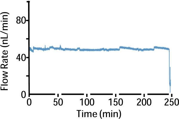
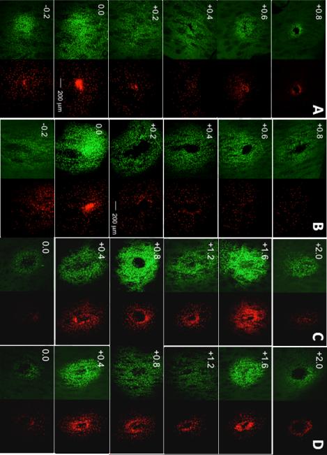
New method: Recently push-pull perfusion at low flow rates (50nL/min) has been proposed as a method for in vivo sampling from the central nervous system. Tissue damage from such probes has not been investigated in detail. In this work, we evaluated acute tissue response to low-flow push-pull perfusion by infusing the nuclear stains Sytox Orange and Hoechst 33342 through probes implanted in the striatum for 200min, to label damaged and total cells, respectively, in situ.

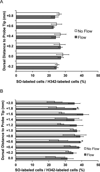
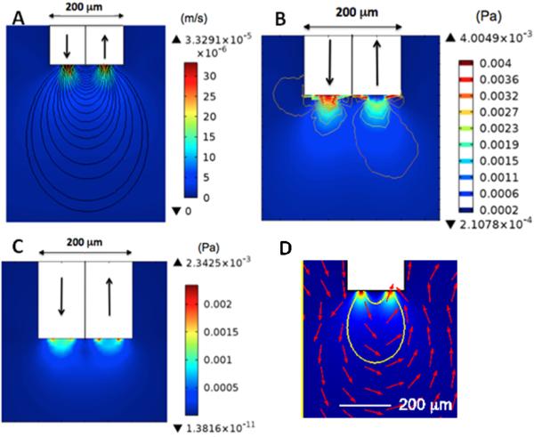
Results: Using the damaged/total labeled cell ratio as a measure of tissue damage, we found that 33±8% were damaged within the dye region around a microdialysis probe. We found that low-flow push-pull perfusion probes damaged 24±4% of cells in the sampling area. Flow had no effect on the number of damaged cells for low-flow push-pull perfusion. Modeling revealed that shear stress and pressure gradients generated by the flow were lower than thresholds expected to cause damage. Comparison with existing methods.Push-pull perfusion caused less tissue damage but yielded 1500-fold better spatial resolution.

Conclusions: Push-pull perfusion at low flow rates is a viable method for sampling from the brain with potential for high temporal and spatial resolution. Tissue damage is mostly caused by probe insertion. Smaller probes may yield even lower damage.

Keywords: Brain tissue damage; Cell viability; Computational modeling; In vivo sampling; Microdialysis; Push–pull perfusion.


Figures

Figure 1
Figure 1
Diagrams and photomicrographs of low-flow PPP (A) and microdialysis (B) probes tested with dye staining experiments. Low-flow PPP probe is oriented with sampling tip down. Arrows illustrate direction of flow.
Figure 2
Figure 2
Sample trace of pull flow rate during low-flow PPP in vivo. Flow rate was recorded by a flow meter as vacuum was used to pull flow. Push flow rate in PPP was maintained at 50 nL/min by a syringe pump to match pull flow rate.
Figure 3
Figure 3
Representative fluorescent confocal microscopy images (voxel size 2.5 × 2.5 × 3.2 μm) of cell viability-stained 100 μm horizontal sections in PPP with no flow (A), PPP with sampling flow of 50 nL/min (B), microdialysis no flow (C), and microdialysis flow (D). Left column for each probe condition shows H342 (pseudo-colored green) staining and the right column shows SO (pseudo-colored red) staining. Stains were infused together at the end of the sampling period to gauge extent of damaged cells (stained by SO) relative to total cells stained (H342). Sections are labeled according to their relative vertical distance from the sampling tip (0.0) in mm with negative values indicating more ventral. All photomicrographs have identical scaling (scale bar = 200 μm).
Figure 4
Figure 4
Fraction of stained cells that were stained with Sytox Orange (SO), a membrane impermeable dye for PPP (A) and microdialysis (B) for flow and no flow conditions. The number of cells stained with H342, a membrane permeable dye, was counted as the total number of stained cells to calculate the fraction. SO labeled cells are considered to be damaged. Sections with significant differences (p < 0.05) in damaged/total cell ratio between flow and non- flow are labeled with an asterisk. Error bars are SEM (n = 4).
Figure 5
Figure 5
The gradient of SO-labeled/H342-labeled cells with respect to the horizontal probe hole center in PPP and microdialysis for flow conditions (n = 4 for each group). Cell count was performed within concentric rings of increasing radii at peak cell count sections: at the probe tip (0.0) in PPP, and +1.0 mm from the probe tip in microdialysis. Significant differences (p < 0.05) between PPP flow and microdialysis flow only existed within 250 μm of the probe hole center. Error bars are SEM. Inset: example of concentric rings used to calculate the gradient of dead/total cell ratio in the transverse plane. The image shown is from a PPP sample.
Figure 6
Figure 6
Numerical modeling of low flow PPP at 50 nL/min. In all drawings, the white area represents the sampling capillary tip and arrows indicate direction of flow. (A) Calculated velocity map. (B) Calculated pressure map. (C) Calculated shear stress map. (D) Close up of data from A with streamlines (in red) indicating direction of flow around probe tip.

Similar articles

Cited by

References

    1. Benveniste H, Drejer J, Schousboe A, Diemer NH. Regional cerebral glucose phosphorylation and blood-flow after insertion of a microdialysis fiber through the dorsal hippocampus in the rat. Journal of Neurochemistry. 1987;49:729–34. - PubMed
    1. Bjornsson CS, Lin G, Al-Kofahi Y, Narayanaswamy A, Smith KL, Shain W, Roysam B. Associative image analysis: A method for automated quantification of 3D multi-parameter images of brain tissue. Journal of Neuroscience Methods. 2008;170:165–78. - PMC - PubMed
    1. Caicedo HH, Hernandez M, Fall CP, Eddington DT. Multiphysics simulation of a microfluidic perfusion chamber for brain slice physiology. Biomedical Microdevices. 2010;12:761–7. - PMC - PubMed
    1. Cellar NA, Kennedy RT. A capillary-PDMS hybrid chip for separations-based sensing of neurotransmitters in vivo. Lab on a Chip. 2006;6:1205–12. - PubMed
    1. Clapp-Lilly KL, Roberts RC, Duffy LK, Irons KP, Hu Y, Drew KL. An ultrastructural analysis of tissue surrounding a microdialysis probe. Journal of Neuroscience Methods. 1999;90:129–42. - PubMed

Publication types

LinkOut - more resources