. 2015 Mar;199(3):777-91.
doi: 10.1534/genetics.115.174300. Epub 2015 Jan 21.

Muscle cell fate choice requires the T-box transcription factor midline in Drosophila

Affiliations

Muscle cell fate choice requires the T-box transcription factor midline in Drosophila

Ram P Kumar et al. Genetics. 2015 Mar.

Abstract

Drosophila Midline (Mid) is an ortholog of vertebrate Tbx20, which plays roles in the developing heart, migrating cranial motor neurons, and endothelial cells. Mid functions in cell-fate specification and differentiation of tissues that include the ectoderm, cardioblasts, neuroblasts, and egg chambers; however, a role in the somatic musculature has not been described. We identified mid in genetic and molecular screens for factors contributing to somatic muscle morphogenesis. Mid is expressed in founder cells (FCs) for several muscle fibers, and functions cooperatively with the T-box protein H15 in lateral oblique muscle 1 and the segment border muscle. Mid is particularly important for the specification and development of the lateral transverse (LT) muscles LT3 and LT4, which arise by asymmetric division of a single muscle progenitor. Mid is expressed in this progenitor and its two sibling FCs, but is maintained only in the LT4 FC. Both muscles were frequently missing in mid mutant embryos, and LT4-associated expression of the transcription factor Krüppel (Kr) was lost. When present, LT4 adopted an LT3-like morphology. Coordinately, mid misexpression caused LT3 to adopt an LT4-like morphology and was associated with ectopic Kr expression. From these data, we concluded that mid functions first in the progenitor to direct development of LT3 and LT4, and later in the FCs to influence whichever of these differentiation profiles is selected. Mid is the first T-box factor shown to influence LT3 and LT4 muscle identity and, along with the T-box protein Optomotor-blind-related-gene 1 (Org-1), is representative of a new class of transcription factors in muscle specification.

Keywords: Tbx20; differentiation; founder cell; midline; muscle identity.


Figures

Figure 1
Figure 1
Identification of mid as a gene regulating somatic muscle morphogenesis. (A) The mid/H15 genomic region and corresponding map positions of deletions used in this study. The minimal overlapping area among the deficiencies that fail to complement contains three protein coding genes, six uncharacterized nonprotein coding genes, and one snoRNA gene (not shown). The molecular breakpoints are shown for deficiencies that have been mapped to genome sequence. (B) The sequence lesion in the nonnull allele mid8A69N1 changes aa 300 in the T-box domain from N to I. (C) Schematic representation of somatic muscle pattern, with analyzed muscles shown in red. (D–M) Stage 16 embryos stained with muscle myosin corresponding to wild-type (D and E), mid1/mid8A69N1 (F and G), nmr1210 (H and I), mid1/Df(2L)BSC810 (J and K), and Df(2L)x528/Df(2L)GpdhA, which lacks both mid and H15 (L and M) embryos. Lateral (D, F, H, J, and L) and ventral (E, G, I, K, and M) views. The arrowhead in F and J denotes the change in position of the fourth LT compared to wild type. The 1–4 represents LT1–4 muscles. Bar, 15 µm.
Figure 2
Figure 2
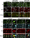
Mid and Kr expression in the muscle lineages. All are lateral views of a single abdominal hemisegment of embryos immunostained for Kr (red, A–G and I–M) and Mid (green A′–G′ and I′–M′), with merged data in (A′′–G′′ and I′′–M′′). (A) Coexpression of Mid and Kr is seen in the LT3/4 and VA1/2 progenitors. (B) By mid stage 11, coexpression of Mid and Kr are observed in FCs for LT3, LT4, VA1, and VA2. (C and I) Mid and Kr coexpression continues in FCs for LT4, VA1, and VA2 in late stage 11, but both Mid and Kr are reduced in the FC for LT3. (D–G) Mid and Kr are lost from the developing LT3 myotube but continue to be detected in LT4. (H) Schematic representation of Mid and Kr coexpression during LT3, LT4, VA1, VA2, and LO1 development. (J) No expression is apparent in FCs for LO1 and VT1 at stage 12, though Mid is apparent in FCs for LT4 and VA2 as in D–G. (K–M) Mid expression is detectable in the LO1 FC and becomes apparent in the muscle by stage 15. The asterisk in A–G and I–M indicates nonmuscle Mid-expressing cells. Where indicated, m and l refer to mid and late stage, respectively. Bar, 10 µm.
Figure 3
Figure 3
Founder cells for LT3, LT4, LO1, and the VT1, but not VA2, are impacted by the loss of mid. Lateral views of abdominal hemisegments from stage 11 embryos (B and C), stage 14 (D and E), and stage 13 (F–I), immunostained for Kr, Slou, and Ara, as indicated. Wild type is shown in B, D–D′′, F–F′′′, and G–G′′′, while Df(2L)x528/Df(2L)GpdhA, which lacks both mid and H15, is shown in C, E–E′′, H–H′′′, and I–I′′′. Asterisks indicate missing FCs for LT3 and LT4 (C and E), and LO1 (E′). The absence of both Kr- and Ara-expressing FCs for LT3 and LT4 was apparent in stage 13 embryos (H–I′′′). Enlarged view of area outlined in white (F and H) is shown in G and I, repectively. Note that expression of Ara in the FC for LO1 has not been reported previously. Bar, 10 µm.
Figure 4
Figure 4
Panmesodermal expression of Mid rescues Kr-expressing cells in mid mutants and generates extra Kr-expressing cells. Lateral views of stage 14 embryos immunostained for Kr. (A) mid1/mid1; Mef2-GAL4/+, (B) mid1, UAS-mid/mid1; Mef2-GAL4/+, (C) UAS-mid/+; Mef2-GAL4/+, (D) 24B-GAL4/+, and (E) UAS-mid/+; 24B-GAL4/+. (A′–F′) Digital 2D snapshots of Kr-expressing cells that were isosurfaced from confocal Z-series in 3D using Imaris (Bitplan). (A) Kr-expressing cells in the position of LT4 were absent in mid mutants, suggesting that mid is critical for specification and/or Kr expression in the FC for LT4. (B) The presence of Kr-expressing cells in the LT position of mid mutant embryos upon panmesodermal expression of mid suggests that mid expression is sufficient for the LT4 FC. (F and F′) Enlarged view from E and E′. (C, E, and F) Additional Kr-expressing cells were visible in the LT position upon misexpression of mid in wild-type embryos (asterisks). Bar, 10 µm.
Figure 5
Figure 5
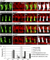
Loss of muscles LT3 and LT4 in mid and mid, H15 mutant embryos. (A–D) Lateral views of stage 16 embryos immunostained for tropomyosin (Tropo) and for GFP driven by the ap-GFP transgene. (A–A′′′) ap-GFP control. (B–B′′′ and D–D′′′) Embryos trans-heterozygous for mid1/Df(2L)BSC810; ap-GFP. (C–C′′′) Embryos trans-heterozygous for Df(2L)x528/Df(2L)GpdhA; ap-GFP that lack mid and H15. (A′′′–D′′′) Digital 2D snapshot of GFP-expressing LTs that were isosurfaced in 3D from confocal Z-series in Imaris (Bitplan). (D′′′) Note small extra syncytia (yellow) in some segments of mid mutant embryos. Arrows indicate LTs and the arrowhead in D refers to the altered ventral limit of LT4. Bar, 15 µm. (E) Quantitation of phenotypes produced by loss of H15 alone, mid alone, mid and H15, or panmesodermal expression of Mid. n = abdominal hemisegments.
Figure 6
Figure 6
Panmesodermal expression of Mid causes a morphological conversion of LT3 to LT4. Lateral views of stage 16 embryos immunostained for tropomyosin (Tropo) and GFP directed by the ap-GFP transgene. (A–A′′′ and C) ap-GFP/24B-GAL4 and (B–B′′′ and D) UAS-mid/+; ap-GFP/24B-GAL4. (A′′′–B′′′) Digital 2D snapshot of GFP-expressing LTs that were isosurfaced in 3D from confocal Z-series in Imaris (Bitplan). (B′′′) Note that additional small syncytia are apparent in some segments (yellow). The asterisk in B denotes segments with extra muscles, arrowhead denotes the dorsally shifted LT3 muscle, and arrows indicate LTs. (C–E) Enlarged views from A′′′, B′′′, and Figure 2D′′′, respectively, showing the dorsal shift of LT4 in wild-type embryos (C), dorsal shift of both LT3 and LT4 upon panmesodermal expression of Mid (D), and lack of this shift in mid mutants (E). Bar, 15 µm. (F) Quantitation of the difference in the ventral limit in wild-type, panmesodermal expression of Mid and loss of mid. LT2/3, LT2–4, and LT3/4 corresponds to the height difference between LT2 and LT3, LT2 and LT4, and LT3 and LT4, respectively. n = abdominal hemisegments.
Figure 7
Figure 7
Loss and gain of Mid affects the number of fusion events in LT3 and LT4. (A–C) Lateral views of stage 17 live embryos expressing the apRed transgene. Approximately six hemisegments are shown. (A) Wild type (apRed) embryo. (B) mid1; apRed homozygote. (C) twi-GAL4, apRed > UAS-Mid. (D) Box and whisker plot showing comparison of the number of apRed-positive nuclei only in the fourth most posterior LT muscle in a hemisegment. Significance (P = 0.03) is indicated with an *. (E) Comparison of the number of apRed-positive nuclei only in the third most posterior LT muscle in a hemisegment. Significance is shown as ****P < 0.0001. (F) Comparison of the number of apRed-positive nuclei per LT muscle in a single hemisegment between wild type and mid1; apRed mutant. Significance is shown as **P = 0.0015. (G) Comparison of the number of apRed-positive nuclei per LT muscle in mid1; apRed mutant hemisegments where there were two, three, or four total LT muscles. Significance between two and three LTs is indicated as **P = 0.0049. Significance between two and four LTs is indicated as ****P = 1.75 × 10⁻⁷. Bar, 25 μm.

Similar articles

Cited by

References

    1. Abmayr S. M., Keller C. A., 1998. Drosophila myogenesis and insights into the role of nautilus. Curr. Top. Dev. Biol. 38: 35–80. - PubMed
    1. Abmayr S. M., Pavlath G. K., 2012. Myoblast fusion: lessons from flies and mice. Development 139: 641–656. - PMC - PubMed
    1. Bataille L., Delon I., Da Ponte J. P., Brown N. H., Jagla K., 2010. Downstream of identity genes: muscle-type-specific regulation of the fusion process. Dev. Cell 19: 317–328. - PMC - PubMed
    1. Bate M., 1990. The embryonic development of larval muscles in Drosophila. Development 110: 791–804. - PubMed
    1. Baylies M. K., Bate M., 1996. twist: a myogenic switch in Drosophila. Science 272: 1481–1484. - PubMed

Publication types

MeSH terms

LinkOut - more resources