. 2015 Apr 10;290(15):9542-54.
doi: 10.1074/jbc.M114.596098. Epub 2015 Jan 22.

Biased signaling at chemokine receptors

Affiliations

Biased signaling at chemokine receptors

Jenny Corbisier et al. J Biol Chem. .

Abstract

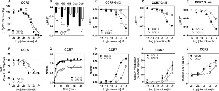
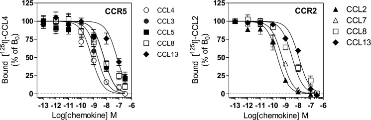
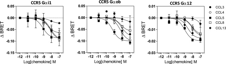
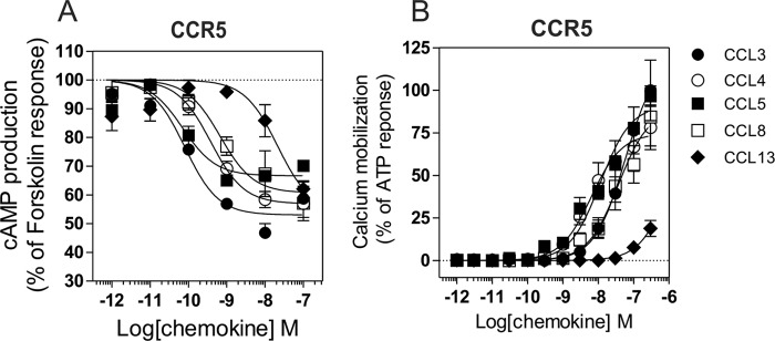
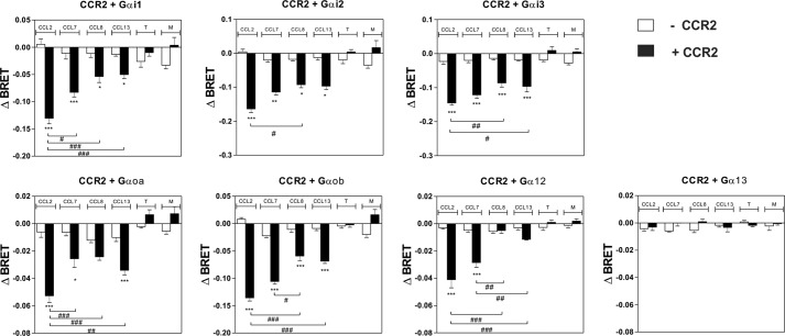
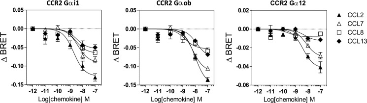
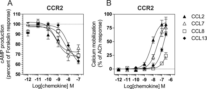
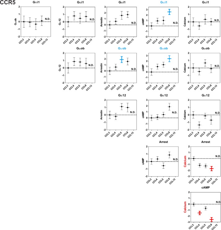
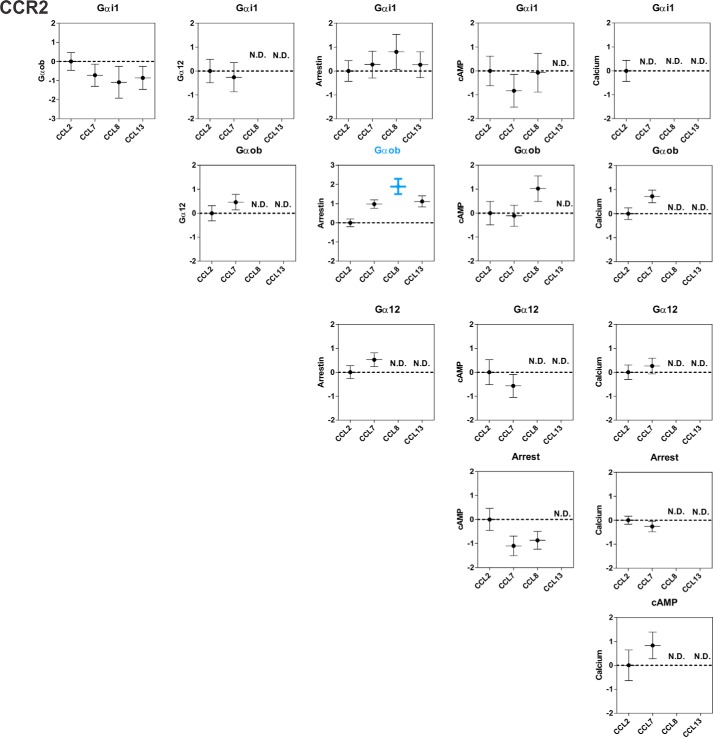
The ability of G protein-coupled receptors (GPCRs) to activate selective signaling pathways according to the conformation stabilized by bound ligands (signaling bias) is a challenging concept in the GPCR field. Signaling bias has been documented for several GPCRs, including chemokine receptors. However, most of these studies examined the global signaling bias between G protein- and arrestin-dependent pathways, leaving unaddressed the potential bias between particular G protein subtypes. Here, we investigated the coupling selectivity of chemokine receptors CCR2, CCR5, and CCR7 in response to various ligands with G protein subtypes by using bioluminescence resonance energy transfer biosensors monitoring directly the activation of G proteins. We also compared data obtained with the G protein biosensors with those obtained with other functional readouts, such as β-arrestin-2 recruitment, cAMP accumulation, and calcium mobilization assays. We showed that the binding of chemokines to CCR2, CCR5, and CCR7 activated the three Gαi subtypes (Gαi1, Gαi2, and Gαi3) and the two Gαo isoforms (Gαoa and Gαob) with potencies that generally correlate to their binding affinities. In addition, we showed that the binding of chemokines to CCR5 and CCR2 also activated Gα12, but not Gα13. For each receptor, we showed that the relative potency of various agonist chemokines was not identical in all assays, supporting the notion that signaling bias exists at chemokine receptors.

Keywords: Bioluminescence Resonance Energy Transfer (BRET); Biosensor; Chemokine; G Protein-coupled Receptor (GPCR); Signaling.


Figures

FIGURE 1.
FIGURE 1.
A, competition binding assays performed on CHO-K1 cells expressing CCR7. Cells were incubated with 0.1 nm 125I-CCL19 as tracer and unlabeled CCL19 (●) or CCL21 (○) as competitors. The data were normalized for nonspecific binding (0%) in the presence of 300 nm of competitor (CCL19) and specific binding in the absence of competitor (100%). All points were run in triplicate (error bars indicate S.E.). B–E, G protein activation by CCR7. Real time measurement of BRET signal in HEK293T cells coexpressing G protein biosensors and CCR7 and stimulated for 1 min with 50 nm CCL19 (black bars and circles) or CCL21 (open bars and circles) is shown. The results are expressed as the differences in BRET signals measured in the presence and absence of stimulation. The data represent the means ± S.E. of three to six independent experiments. Statistical significance between stimulated and unstimulated cells was assessed using Tukey's test. ***, p < 0.001. F, inhibition of cAMP by CCR7. Measurement of BRET signal in HEK293T cells coexpressing cAMP biosensor and CCR7 and stimulated sequentially by CCL19 (●) or CCL21 (○) and forskolin. The results were normalized for the basal signal in absence of stimulation (0%), and the maximal response was obtained with forskolin only (100%). The data represent the means ± S.E. of three independent experiments. G and H, recruitment of β-arrestin 2 by CCR7. Real time measurement of BRET signal in HEK293T cells expressing β-arrestine2-RLuc8 and CCR7-Venus and stimulated with CCL19 (black circles) or CCL21 (open circles) is shown. Cells were stimulated by 100 nm chemokines in E and for 30 min in F. The results are expressed as the difference in BRET signals measured in the presence and absence of stimulation. The data represent the means ± S.E. of three independent experiments. I, calcium mobilization by CCR7. Calcium mobilization was measured in HEK293 cells using the aequorin-based functional assay. Cells expressing CCR7 were stimulated with increasing concentrations of CCL19 (●) or CCL21 (○), and luminescence was recorded for 30 s. The results were normalized for the basal luminescence in absence of agonist (0%), and the maximal response was obtained with 50 μm acetylcholine (100%). The data represent the means ± S.E. of three independent experiments. J, phosphorylation of ERK1/2 by CCR7. HEK293 cells stably expressing CCR7 were stimulated with 100 nm CCL19 or CCL21 for 2 min. The results are expressed as the ratio between the amount of phospho-ERK1/2 and total ERK1/2 following quantification on Western blots. The data represent the means ± S.E. of three independent experiments.
FIGURE 2.
FIGURE 2.
A, competition binding assays performed on cells expressing CCR5. CHO-K1 cells expressing CCR5 were incubated with 0.1 nm 125I-CCL4 as tracer and unlabeled CCL3, CCL4, CCL5, CCL8, and CCL13 as competitors. The data were normalized for nonspecific binding (0%) in the presence of 300 nm CCL4 and specific binding in the absence of competitor (100%). All points were run in triplicate (error bars indicate S.E.). B, competition binding assays performed on cells expressing CCR2. CHO-K1 cells expressing CCR2 were incubated with 0.05 nm 125I-CCL2 as tracer and unlabeled CCL5, CCL7, CCL8, and CCL13 as competitors. The data were normalized for nonspecific binding (0%) in the presence of 300 nm CCL2 and specific binding in the absence of competitor (100%). All points were run in triplicate (error bars indicate S.E.).
FIGURE 3.
FIGURE 3.
Panel of G proteins activated by CCR5. Real time measurement of BRET signal in HEK293T cells coexpressing CCR5 and G protein biosensors (black bars) or G protein biosensors only (open bars) and stimulated for 1 min with 100 nm of the chemokines CCL3, CCL4, CCL5, CCL7, CCL8, and CCL13 or 1 μm of the small molecules TAK779 (T) and Maraviroc (M) is shown. The results are expressed as the difference in BRET signal measured in the presence and absence of stimulation. The data represent the means ± S.E. of at least six independent experiments. Statistical significance between cells expressing or not CCR5. (***, p < 0.001; **, p < 0.01; *, p < 0.1) and between chemokines (###, p < 0.001; ##, p < 0.01; #, p < 0.1) was assessed using Tukey's test.
FIGURE 4.
FIGURE 4.
Activation of Gi1, Gob, and G12 by CCR5. Real time measurement of BRET signal in HEK293T cells coexpressing CCR5 and G protein biosensors and stimulated with increasing concentration of chemokines is shown. The results are expressed as the differences in BRET signal measured in the presence and absence of chemokines. The data represent the means ± S.E. of three independent experiments.
FIGURE 5.
FIGURE 5.
Inhibition of cAMP and calcium mobilization triggered by CCR5. A, measurement of BRET signal in HEK293T cells coexpressing the cAMP biosensor and CCR5 and stimulated sequentially by chemokines and forskolin. The results were normalized for the basal signal in absence of stimulation (0%), and the maximal response was obtained with forskolin only (100%). The data represent the means ± S.E. of three independent experiments. B, calcium mobilization was measured in CHO-K1 cells using the aequorin-based functional assay. Cells expressing CCR5 were stimulated with increasing concentrations of chemokines, and luminescence was recorded for 30 s. The results were normalized for the basal luminescence in absence of agonist (0%), and the maximal response was obtained with 25 μm ATP (100%). The data represent the means ± S.E. of three independent experiments.
FIGURE 6.
FIGURE 6.
Panel of G proteins activated by CCR2. Real time measurement of BRET signal in HEK293T cells coexpressing CCR2 and G protein biosensors (black bars) or G protein biosensors only (open bars) and stimulated for 1 min with 100 nm of the chemokines CCL2, CCL7, CCL8, and CCL13 or 1 μm of the small molecules TAK779 (T) and Maraviroc (M) is shown. The results are expressed as the difference in BRET signal measured in the presence and absence of stimulation. The data represent the means ± S.E. of at least six independent experiments. Statistical significance between cells expressing or not CCR2 (***, p < 0.001; **, p < 0.01; *, p < 0.1) and between chemokines (###, p < 0.001; ##, p < 0.01; #, p < 0.1) was assessed using Tukey's test.
FIGURE 7.
FIGURE 7.
Activation of Gi1, Gob, and G12 by CCR2. Real time measurement of BRET signal in HEK293T cells coexpressing CCR2 and G protein biosensors and stimulated with increasing concentrations of chemokines is shown. The results are expressed as the differences in BRET signal measured in the presence and absence of chemokines. The data represent the means ± S.E. of three independent experiments.
FIGURE 8.
FIGURE 8.
Inhibition of cAMP and calcium mobilization triggered by CCR2. A, measurement of BRET signal in HEK293T cells coexpressing the cAMP biosensor and CCR2 and stimulated sequentially by chemokines and forskolin. The results were normalized for the basal signal in absence of stimulation (0%), and the maximal response was obtained with forskolin only (100%). The data represent the means ± S.E. of three independent experiments. B, calcium mobilization was measured in HEK293 cells using the aequorin-based functional assay. Cells expressing CCR2 were stimulated with increasing concentrations of chemokines and luminescence was recorded for 30 s. The results were normalized for the basal luminescence in absence of agonist (0%), and the maximal response was obtained with 50 μm acetylcholine (100%). The data represent the means ± S.E. of three independent experiments.
FIGURE 9.
FIGURE 9.
Recruitment of β-arrestin 2 by CCR5 and CCR2. A–D, real time measurement of BRET signal in HEK293T cells expressing either β-arrestin2-RLuc8 and CCR5-Venus (A and B) or β-arrestin2-RLuc8 and CCR2-Venus (C and D) and stimulated with chemokines. For kinetics, BRET signals were measured after addition of 100 nm chemokines. For dose-response curves, BRET was recorded 30 min after stimulation with various concentrations of chemokines. The results are expressed as net BRET, corresponding to the difference in BRET signal between cells expressing arrestin plus receptor and cells expressing arrestin only. The data represent the means ± S.E. of three independent experiments.
FIGURE 10.
FIGURE 10.
Chemokine bias factors at CCR5. Bias factors between different pathways were calculated for each chemokine, using CCL3 as the reference chemokine. Bias factors that are significantly different (Tukey's test) from the reference chemokine CCL3 are in bold and colored according to the preferred pathway. The data represent the means ± S.E. from Table 4. ND, not determined.
FIGURE 11.
FIGURE 11.
Chemokine bias factors at CCR2. Bias factor between different pathways were calculated for each chemokine, using CCL2 as the reference chemokine. Bias factors that are significantly different (Tukey's test) from the reference are in bold and colored according to the preferred pathway. The data represent the means ± S.E. from Table 4. ND, not determined.

References

    1. Lagerström M. C., Schiöth H. B. (2008) Structural diversity of G protein-coupled receptors and significance for drug discovery. Nat. Rev. Drug. Discov. 7, 339–357 - PubMed
    1. Strange P. G. (2008) Signaling mechanisms of GPCR ligands. Curr. Opin. Drug. Discov. Devel. 11, 196–202 - PubMed
    1. Gether U. (2000) Uncovering molecular mechanisms involved in activation of G protein-coupled receptors. Endocr. Rev. 21, 90–113 - PubMed
    1. Marrari Y., Crouthamel M., Irannejad R., Wedegaertner P. B. (2007) Assembly and trafficking of heterotrimeric G proteins. Biochemistry 46, 7665–7677 - PMC - PubMed
    1. Rajagopal S., Rajagopal K., Lefkowitz R. J. (2010) Teaching old receptors new tricks: biasing seven-transmembrane receptors. Nat. Rev. Drug. Discov. 9, 373–386 - PMC - PubMed

Publication types

MeSH terms