

Cor triatriatum sinister identified after new onset atrial fibrillation in an elderly man
- PMID: 25614746
- PMCID: PMC4295417
- DOI: 10.1155/2014/674018
Cor triatriatum sinister identified after new onset atrial fibrillation in an elderly man
Abstract
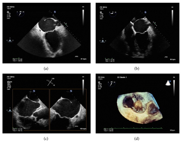
A 73-year-old man with new onset atrial fibrillation with rapid ventricular response underwent transthoracic echocardiography that revealed an echogenic linear structure along the left atrium, suggestive of cor triatriatum sinister (CTS). CTS was confirmed with transesophageal echocardiography which demonstrated a proximal accessory atrium receiving pulmonary venous flow separated from a distal true atrium by a fibromuscular membrane with a large fenestration allowing flow between the chambers. In CTS, the left atrium is divided into proximal and distal chambers by a fenestrated fibromuscular septum. This cardiac anomaly accounts for 0.1% of cases of congenital heart disease and rarely presents in adults. CTS is primarily diagnosed with echocardiography and is associated with left atrial enlargement and development of atrial fibrillation. Treatment options depend on size of the communication between proximal and distal chambers, the gradient across the membrane, and the position of pulmonary veins. In some instances, surgical resection of the membrane that divides the left atrium is warranted.
Figures


References
-
- Richardson J. V., Doty D. B., Siewers R. D., et al. Cor triatriatum (subdivided left atrium) The Journal of Thoracic and Cardiovascular Surgery. 1981;81(2):232–238. - PubMed
-
- Church W. S. Congenital malformation of heart: abnormal septum in the left auricle. Transactions of the Pathological Society of London. 1868;19:188–190.
LinkOut - more resources
Full Text Sources
Other Literature Sources
Research Materials
