. 2015 Jan 1;71(Pt 1):127-35.
doi: 10.1107/S1399004714025280. Epub 2015 Jan 1.

A national facility for biological cryo-electron microscopy

Affiliations

A national facility for biological cryo-electron microscopy

Helen R Saibil et al. Acta Crystallogr D Biol Crystallogr. .

Abstract

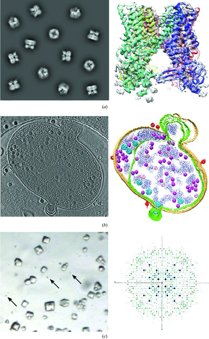
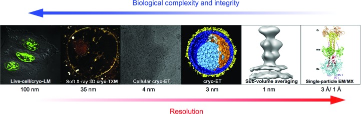
Three-dimensional electron microscopy is an enormously powerful tool for structural biologists. It is now able to provide an understanding of the molecular machinery of cells, disease processes and the actions of pathogenic organisms from atomic detail through to the cellular context. However, cutting-edge research in this field requires very substantial resources for equipment, infrastructure and expertise. Here, a brief overview is provided of the plans for a UK national three-dimensional electron-microscopy facility for integrated structural biology to enable internationally leading research on the machinery of life. State-of-the-art equipment operated with expert support will be provided, optimized for both atomic-level single-particle analysis of purified macromolecules and complexes and for tomography of cell sections. The access to and organization of the facility will be modelled on the highly successful macromolecular crystallography (MX) synchrotron beamlines, and will be embedded at the Diamond Light Source, facilitating the development of user-friendly workflows providing near-real-time experimental feedback.

Keywords: three-dimensional electron microscopy.


Figures

Figure 1
Figure 1
Overview of current methods in biological cryo-EM. (a) Single-particle reconstruction of the TrpV channel (Liao et al., 2013; EMD-5778). Left, projected views representing idealized single-particle data. Right, three-dimensional reconstruction of the mainly α-helical tetramer, coloured by subunit, with the fitted secondary structure. (b) Zernicke phase-contrast cryo-ET of a virus-infected cyanobacterial cell. Left, section through the tomogram; right, segmented view of the cell with the viruses in pink. Reproduced by permission from Macmillan Publishers Ltd, Dai et al. (2013 ▶), copyright (2013). (c) Electron diffraction of lysozyme microcrystals. Left, optical micrograph of small crystals with microcrystals indicated by arrows. Right, representation of the three-dimensional electron diffraction data. Figures reproduced or modified from Shi et al. (2013 ▶) under a Creative Commons Attribution license.
Figure 2
Figure 2
Dynamics of GroEL–ATP (Clare et al., 2012 ▶). Cryo-EM maps (transparent surfaces) and flexible fitting show some of the main structural states determined by multivariate statistical analysis of a 60 000-particle data set. Helices H and I, which denote the hydrophobic binding sites for non-native proteins, are shown in orange/red and the GroES lid is shown in green. (a) Apo GroEL, (b) GroEL–ATP7, Rs1 state, (c) GroEL–ATP7, Rs-open state, (d) GroEL–GroES–ATP.
Figure 3
Figure 3
Integrated structural biology approach combining correlative light microscopy, soft X-ray cryo-microscopy and electron cryo-microscopy with high-resolution structure information, thus enabling the dissection of dynamic processes at different levels of resolution and complexity. The biological process is visualized by light microscopy (LM), transmission X-ray cryo-microscopy (cryo-TXM), electron cryo-tomography (cryo-ET), single-particle cryo-EM and/or macromolecular crystallography (MX). Adapted from Zeev-Ben-Mordehai et al. (2014 ▶).

References

    1. Al-Amoudi, A., Díez, D. C., Betts, M. J. & Frangakis, A. S. (2007). Nature (London), 450, 832–837. - PubMed
    1. Amunts, A., Brown, A., Bai, X.-C., Llácer, J. L., Hussain, T., Emsley, P., Long, F., Murshudov, G., Scheres, S. H. W. & Ramakrishnan, V. (2014). Science, 343, 1485–1489. - PMC - PubMed
    1. Bartesaghi, A., Lecumberry, F., Sapiro, G. & Subramaniam, S. (2012). Structure, 20, 2003–2013. - PMC - PubMed
    1. Chang, Y.-W., Chen, Y., Tocheva, E. I., Treuner-Lange, A., Löbach, S., Søgaard-Andersen, L. & Jensen, G. (2014). Nature Methods, 11, 737–739. - PMC - PubMed
    1. Chen, S., McMullan, G., Faruqi, A. R., Murshudov, G. N., Short, J. M., Scheres, S. H. W. & Henderson, R. (2013). Ultramicroscopy, 135, 24–35. - PMC - PubMed

Publication types