Review
. 2015 Jan 1;71(Pt 1):3-18.
doi: 10.1107/S2053230X14026843. Epub 2015 Jan 1.

A comprehensive review of the lipid cubic phase or in meso method for crystallizing membrane and soluble proteins and complexes

Affiliations
Review

A comprehensive review of the lipid cubic phase or in meso method for crystallizing membrane and soluble proteins and complexes

Martin Caffrey. Acta Crystallogr F Struct Biol Commun. .

Abstract

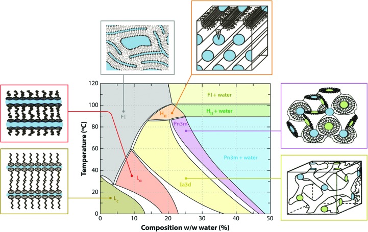
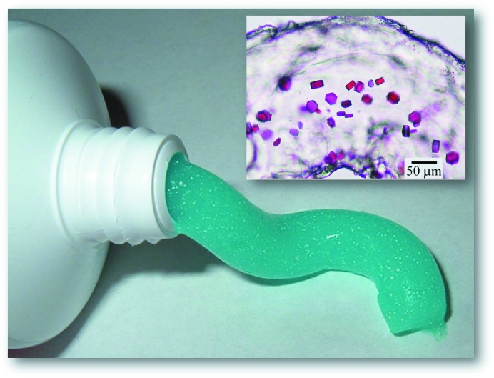
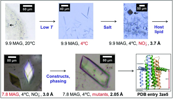
The lipid cubic phase or in meso method is a robust approach for crystallizing membrane proteins for structure determination. The uptake of the method is such that it is experiencing what can only be described as explosive growth. This timely, comprehensive and up-to-date review introduces the reader to the practice of in meso crystallogenesis, to the associated challenges and to their solutions. A model of how crystallization comes about mechanistically is presented for a more rational approach to crystallization. The possible involvement of the lamellar and inverted hexagonal phases in crystallogenesis and the application of the method to water-soluble, monotopic and lipid-anchored proteins are addressed. How to set up trials manually and automatically with a robot is introduced with reference to open-access online videos that provide a practical guide to all aspects of the method. These range from protein reconstitution to crystal harvesting from the hosting mesophase, which is noted for its viscosity and stickiness. The sponge phase, as an alternative medium in which to perform crystallization, is described. The compatibility of the method with additive lipids, detergents, precipitant-screen components and materials carried along with the protein such as denaturants and reducing agents is considered. The powerful host and additive lipid-screening strategies are described along with how samples that have low protein concentration and cell-free expressed protein can be used. Assaying the protein reconstituted in the bilayer of the cubic phase for function is an important element of quality control and is detailed. Host lipid design for crystallization at low temperatures and for large proteins and complexes is outlined. Experimental phasing by heavy-atom derivatization, soaking or co-crystallization is routine and the approaches that have been implemented to date are described. An overview and a breakdown by family and function of the close to 200 published structures that have been obtained using in meso-grown crystals are given. Recommendations for conducting the screening process to give a more productive outcome are summarized. The fact that the in meso method also works with soluble proteins should not be overlooked. Recent applications of the method for in situ serial crystallography at X-ray free-electron lasers and synchrotrons are described. The review ends with a view to the future and to the bright prospects for the method, which continues to contribute to our understanding of the molecular mechanisms of some of nature's most valued proteinaceous robots.

Keywords: crystallization; lipid cubic phase; macromolecular crystallography; membrane-protein structure; mesophase; robot; structure–function; water-soluble proteins.


Figures

Figure 1
Figure 1
Distribution by biological function or activity of integral membrane proteins and peptides crystallized by the in meso method that have yielded crystal structures and records in the Protein Data Bank. The data correspond to the entries in Table 1 ▶ and were sourced from the Protein Data Bank in September 2014.
Figure 2
Figure 2
Annual cumulative number of released PDB records for integral membrane-protein and peptide structures solved with crystals grown by the in meso method. The number of records released each year is indicated. The figure for 2014 is estimated based on a count of 32 recorded up until September 2014. The line is drawn to guide the eye and takes the form y = 5.13exp(0.22x).
Figure 3
Figure 3
Cartoon representation of the events proposed to take place during the crystallization of an integral membrane protein from the lipid cubic mesophase. The process begins with the protein reconstituted into the curved bilayer of the ‘bicontinuous’ cubic phase (tan). Added ‘precipitants’ shift the equilibrium away from stability in the cubic membrane. This leads to phase separation, wherein protein molecules (a) diffuse from the bicontinuous bilayered reservoir of the cubic phase into a sheet-like or lamellar domain and (b) locally concentrate therein in a process that progresses to nucleation and crystal growth. Cocrystallization of the protein with native lipid (cholesterol) is shown in this illustration. As much as possible, the dimensions of the lipid (tan oval with tail), detergent (pink oval with tail), cholesterol (purple), protein (blue and green; β2-adrenergic receptor-T4 lysozyme fusion; PDB entry 2rh1), bilayer and aqueous channels (dark blue) have been drawn to scale. The lipid bilayer is ∼40 Å thick. An expanded view of the various components in the system is shown in (c). Reprinted from Li, Shah et al. (2013 ▶). Copyright 2013 American Chemical Society.
Figure 4
Figure 4
Setting up an in meso crystallization trial manually involves (a) placing membrane-protein solution and lipid into separate gas-tight micro-syringes (typically 50 or 100 µl) connected by a narrow-bore coupler, (b) passing the protein solution and lipid from one syringe to the other via the coupler to effect mixing, homogenization and spontaneous self-assembly of the cubic phase into the bilayer of which the protein has become reconstituted, (c) transferring the optically clear mesophase into one of the syringes, (d) replacing the empty syringe with a dispensing micro-syringe (typically 10 µl) mounted in a repeat dispenser and transferring protein-laden mesophase from the large to the small syringe by way of the coupler, (e) dispensing mesophase followed by precipitant solution into the wells of a glass sandwich crystallization plate and (f) sealing the wells with a glass cover slide. The remaining wells on the plate are filled and sealed and the plate is then incubated at the desired temperature to allow crystallization to occur. An open-access online video of the entire procedure is available (Caffrey & Porter, 2010 ▶).
Figure 5
Figure 5
Temperature–composition phase diagram of the monoolein–water system determined under conditions of use in the heating and cooling directions from 20°C. A schematic representation of the various phase states is included, in which coloured zones represent water. The liquid crystalline phases below ∼17°C are undercooled and metastable (Qiu & Caffrey, 2000 ▶). Abbreviations: FI, fluid isotropic phase; HII, inverted hexagonal phase; Lα, lamellar liquid crystalline phase; Lc, lamellar crystal phase. Reprinted with permission from Caffrey (2009 ▶). Copyright (2009) Annual Reviews.
Figure 6
Figure 6
The cubic phase is viscous and sticky; it has the consistency of thick toothpaste. The particular toothpaste used in this figure contains small white crystals. This is exactly what is sought when in meso crystallization trials are set up. The inset shows crystals of the integral membrane light-driven proton pump bacteriorhodopsin growing in the lipid cubic phase. The analogy between the toothpaste and the crystal-laden mesophase is obvious.
Figure 7
Figure 7
Critical steps in determining the in meso crystal structure of diacylglycerol kinase (DgkA). Diffracting crystals were obtained following extensive temperature, salt and host lipid screening (Li, Shah et al., 2013 ▶). Experimental phasing proved challenging, but finally yielded a structure to a resolution of 2.05 Å (Li et al., 2015 ▶).

References

    1. Aherne, M., Lyons, J. A. & Caffrey, M. (2012). A fast, simple and robust protocol for growing crystals in the lipidic cubic phase. J. Appl. Cryst. 45, 1330–1333. - PMC - PubMed
    1. Ai, X. & Caffrey, M. (2000). Membrane protein crystallization in lipidic mesophases: detergent effects. Biophys. J. 79, 394–405. - PMC - PubMed
    1. Axford, D. et al. (2012). In situ macromolecular crystallography using microbeams. Acta Cryst. D68, 592–600. - PMC - PubMed
    1. Barends, T. R., Foucar, L., Botha, S., Doak, R. B., Shoeman, R. L., Nass, K., Koglin, J. E., Williams, G. J., Boutet, S., Messerschmidt, M. & Schlichting, I. (2014). De novo protein crystal structure determination from X-ray free-electron laser data. Nature (London), 505, 244–247. - PubMed
    1. Berman, H. M., Henrick, K. & Nakamura, H. (2003). Announcing the Worldwide Protein Data Bank (2003). Nature Struct. Biol. 10, 1203–980. - PubMed

Publication types