. 2015 Jan 22;57(2):329-340.
doi: 10.1016/j.molcel.2014.12.028.

A combined proteomics/genomics approach links hepatitis C virus infection with nonsense-mediated mRNA decay

Affiliations

A combined proteomics/genomics approach links hepatitis C virus infection with nonsense-mediated mRNA decay

Holly R Ramage et al. Mol Cell. .

Abstract

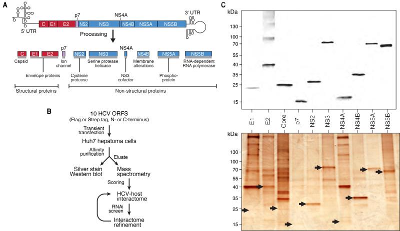
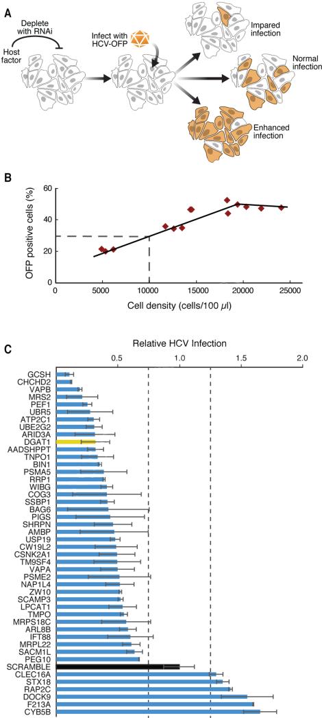
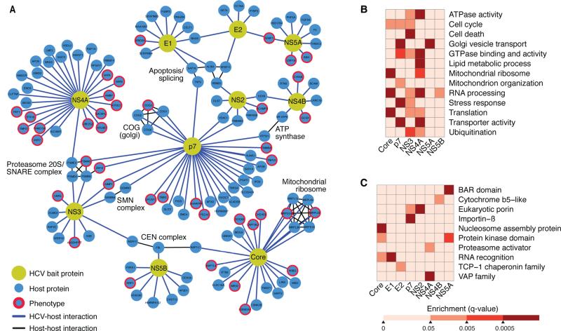
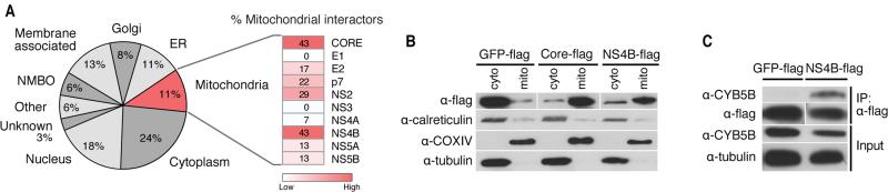
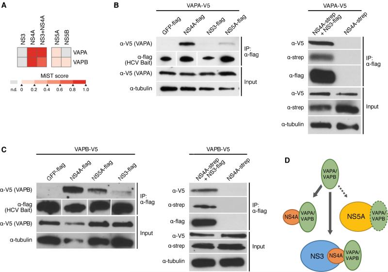
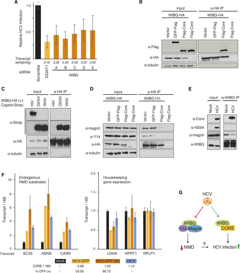
Hepatitis C virus (HCV) is a leading cause of liver disease, but insight into virus-host interactions remains limited. We systematically used affinity purification/mass spectrometry to define the host interactions of all ten HCV proteins in hepatoma cells. We combined these studies with RNAi knockdown of corresponding genes using a two-step scoring approach to generate a map of 139 high-confidence HCV-host protein-protein interactions. We found mitochondrial proteins highly involved in HCV infection and characterized an interaction between the viral core protein and host protein within bgcn homolog (WIBG). Expression of core prevents WIBG from binding its regular interaction partners Y14 and Magoh, two known mediators of the nonsense-mediated mRNA decay pathway. We discovered that this surveillance pathway is disrupted in HCV-infected cells, causing potentially harmful transcripts to accumulate. Our study provides a comprehensive view of HCV-host interactions and uncovers mechanisms for how HCV perturbs host functions during infection.


Figures

Figure 1
Figure 1. Affinity-purification of HCV proteins and interactome strategy
(A) Schematic of HCV genome and 10 proteins derived from the viral polyprotein. (B) Flowchart of the strategy to generate the HCV-host protein-protein interactome. (C) Anti-flag western blot (upper) and silver staining (lower) of 10 flag-tagged HCV proteins after transient transfection and affinity purification from Huh7 cells. Silver-stained bands corresponding to HCV bait proteins are indicated by arrows. See also Figure S1 and Tables S1, S2, S3.
Figure 2
Figure 2. A lentiviral shRNA screen identifies new HCV cofactors
(A) Schematic of the HCV infection assay with an HCV-OFP (orange fluorescent protein mKO2) reporter virus. (B) For each infection, we generated a standard curve of expected infection rates in scrambled shRNA-treated cells by plating the cells at indicated densities and obtaining the percentage of OFP-positive cells. (C) Results of the HCV infection screen. Interacting host proteins that caused changes in infection levels are indicated on the y-axis. Relative infection levels (normalized to cell density) are indicated on the x-axis. The positive control, DGAT1, is shown in yellow, and the negative control, scrambled, in black. Results are shown as average (±SD) of a minimum of six experiments with two independent shRNAs (Experimental Procedures, S5). See also Figure S2 and Table S4.
Figure 3
Figure 3. A complete HCV-host protein-protein interaction network in hepatoma cells
(A) Network representation of the HCV-host interactome in Huh7 cells generated with Cytoscape. There are 10 HCV bait proteins (yellow nodes) and 134 interacting host proteins (blue nodes). Interactions of HCV and host proteins are indicated by blue lines. Curated host-host protein interactions from the CORUM database are indicated by black lines. (B–C) Heat maps representing manually curated enriched biological processes, molecular functions, and pathways (B) and protein domains (C) of the host factors that interact with HCV bait proteins. Colors represent statistical significance (q value) as indicated by the accompanying scale. See also Table S6.
Figure 4
Figure 4. Cellular localization of HCV-interacting host proteins
(A) Localization of HCV-interacting host proteins as a pie chart. Each category contains the percentage of interactors of the total refined interactome that localize to a cell compartment. The heat map indicates the percentage of mitochondrial proteins within each individual bait protein's interactome. (B) Differential centrifugation of HEK293T cells expressing GFP-flag, flag-core, or NS4B-flag to examine protein localization to mitochondria or cytosolic fractions. Control antibodies recognize calreticulin (endoplasmic reticulum), tubulin (cytosol) and COXIV (mitochondria). (C) Immunoprecipitations with Huh7 cell lysates expressing GFP-flag or NS4B-flag were performed with α-flag affinity resin and blotted with the indicated antibodies to demonstrate that NS4B specifically interacts with endogenous CYB5B, a mitochondrial outer membrane protein. See also Figure S3.
Figure 5
Figure 5. HCV NS4A interacts with host proteins VAPA and VAPB
(A) Heat map indicating the interaction scores of VAPA or VAPB with NS3, NS4A, NS3+NS4A, NS5A, and NS5B, as identified by AP-MS. MIST scores in the color scale represent the confidence in the observed interaction. (B) Immunoprecipitations with α-flag resin of Huh7 cell lysates expressing the indicated HCV bait proteins, or GFP as a control, and VAPA. VAPA-V5 interacts strongly with NS4A-flag, weakly with NS5A-flag, and very little with NS3-flag (left). V5-VAPA interacts with NS3-flag when NS4A-strep is coexpressed (right). Results are representative of three independent experiments. (C) Same experiment as in (B) only with VAPB-V5. Results are representative of three independent experiments. (D) Model of the interaction of VAPA and VAPB with NS4A alone, or while NS4A is bound to NS3. See also Figures S4, S5.
Figure 6
Figure 6. HCV infection requires WIBG and disrupts the host nonsense-mediated decay surveillance pathway
(A) Relative infection rates (Figure 2A, B) after knock-down of WIBG with five shRNAs or shRNA targeting DGAT1 or scrambled shRNA as positive and negative controls, respectively. Shown is the average (±SD) of at least 12 replicates in two independent experiments. (B) and (D) Immunoprecipitations in HEK293T cells expressing the indicated constructs along with the flag-tagged HCV core from genotype 2a. Results are representative of three independent experiments. (C) Immunoprecipitations in HEK293T cells expressing WIBG-HA along with the indicated viral capsids from dengue virus, West Nile virus or HIV-1. Results are representative of three independent experiments. (E) Immunoprecipitations of endogenous WIBG in mock- and HCV-OFP SI-infected Huh7.5 cells. Results are representative of two independent experiments. (F) Huh7.5 cells were infected with the indicated HCV constructs. Three endogenous NMD substrate transcripts y (left) and transcripts of three housekeeping genes (right) were analyzed by quantitative RT-PCR. Shown is a representative of three independent experiments. Infection rates based on OFP expression or on quantitative RT-PCR are indicated below. (G) Model of the WIBG-HCV interaction. See text for details. See also Figure S6.

References

    1. Aweya JJ, Tan YJ. Modulation of programmed cell death pathways by the hepatitis C virus. Front Biosci (Landmark Ed) 2011;16:608–618. - PubMed
    1. Balistreri G, Horvath P, Schweingruber C, Zund D, McInerney G, Merits A, Muhlemann O, Azzalin C, Helenius A. The host nonsense-mediated mRNA decay pathway restricts Mammalian RNA virus replication. Cell Host Microbe. 2014;16:403–411. - PubMed
    1. Bartenschlager R, Lohmann V, Wilkinson T, Koch JO. Complex formation between the NS3 serine-type proteinase of the hepatitis C virus and NS4A and its importance for polyprotein maturation. J Virol. 1995;69:7519–7528. - PMC - PubMed
    1. Blight KJ, McKeating JA, Rice CM. Highly permissive cell lines for subgenomic and genomic hepatitis C virus RNA replication. J Virol. 2002;76:13001–13014. - PMC - PubMed
    1. Bono F, Ebert J, Unterholzner L, Guttler T, Izaurralde E, Conti E. Molecular insights into the interaction of PYM with the Mago-Y14 core of the exon junction complex. EMBO Rep. 2004;5:304–310. - PMC - PubMed

Publication types

MeSH terms