

Airway epithelial NF-κB activation promotes the ability to overcome inhalational antigen tolerance
- PMID: 25616105
- PMCID: PMC4472492
- DOI: 10.1111/cea.12491
Airway epithelial NF-κB activation promotes the ability to overcome inhalational antigen tolerance
Abstract
Background: Inhalational antigen tolerance typically protects against the development of allergic airway disease but may be overcome to induce allergic sensitization preceding the development of asthma.
Objectives: We examined in vivo whether pre-existing inhalational antigen tolerance could be overcome by activation of the transcription factor NF-κB in conducting airway epithelial cells, and used a combination of in vivo and in vitro approaches to examine the mechanisms involved.
Methods: Wild-type and transgenic mice capable of expressing constitutively active IκB kinase β (CAIKKβ) in airway epithelium were tolerized to inhaled ovalbumin. Twenty-eight days later, the transgene was transiently expressed and mice were exposed to inhaled OVA on Day 30 in an attempt to overcome inhalational tolerance.
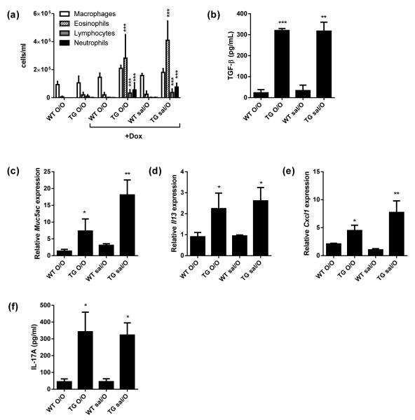
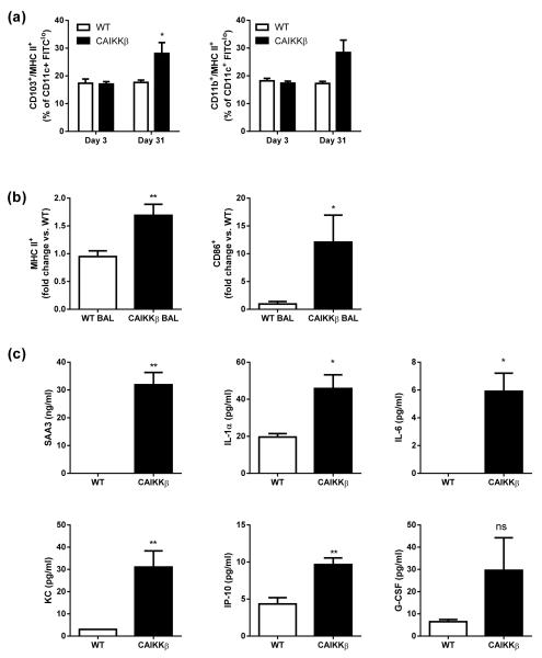
Results: Following ovalbumin challenge on days 40-42, CAIKKβ mice in which the transgene had been activated exhibited characteristic features of allergic airway disease, including airway eosinophilia and methacholine hyper-responsiveness. Increases in the CD103(+) and CD11b(HI) lung dendritic cell populations were present in CAIKKβ mice on Day 31. Bronchoalveolar lavage from mice expressing CAIKKβ mice induced CD4(+) T cells to secrete T(H)2 and T(H)17 cytokines, an effect that required IL-4 and IL-1 signalling, respectively. CAIKKβ mice on Dox demonstrated increased numbers of innate lymphoid type 2 cells (ILC2) in the lung, which also exhibited elevated mRNA expression of the T(H)2-polarizing cytokine IL-4. Finally, airway epithelial NF-kB activation induced allergic sensitization in CAIKKβ mice on Dox that required IL-4 and IL-1 signalling in vivo.
Conclusions: Our studies demonstrate that soluble mediators generated in response to airway epithelial NF-κB activation orchestrate the breaking of inhalational tolerance and allergic antigen sensitization through the effects of soluble mediators, including IL-1 and IL-4, on pulmonary dendritic cells as well as innate lymphoid and CD4(+) T cells.
Keywords: NF-κB; T regulatory cells; allergic airway disease; dendritic cells; inhalational antigen tolerance.
© 2015 John Wiley & Sons Ltd.
Figures


Similar articles
-
Airway epithelial NF-κB activation promotes allergic sensitization to an innocuous inhaled antigen.Am J Respir Cell Mol Biol. 2011 May;44(5):631-8. doi: 10.1165/rcmb.2010-0106OC. Epub 2010 Jun 25. Am J Respir Cell Mol Biol. 2011. PMID: 20581095 Free PMC article.
-
NO2 inhalation induces maturation of pulmonary CD11c+ cells that promote antigenspecific CD4+ T cell polarization.Respir Res. 2010 Jul 26;11(1):102. doi: 10.1186/1465-9921-11-102. Respir Res. 2010. PMID: 20659336 Free PMC article.
-
Restricted aeroallergen access to airway mucosal dendritic cells in vivo limits allergen-specific CD4+ T cell proliferation during the induction of inhalation tolerance.J Immunol. 2011 Nov 1;187(9):4561-70. doi: 10.4049/jimmunol.1004189. Epub 2011 Sep 19. J Immunol. 2011. PMID: 21930961
-
Allergens and the airway epithelium response: gateway to allergic sensitization.J Allergy Clin Immunol. 2014 Sep;134(3):499-507. doi: 10.1016/j.jaci.2014.06.036. J Allergy Clin Immunol. 2014. PMID: 25171864 Review.
-
Type 2 innate lymphoid cells: at the cross-roads in allergic asthma.Semin Immunopathol. 2016 Jul;38(4):483-96. doi: 10.1007/s00281-016-0556-2. Epub 2016 Mar 10. Semin Immunopathol. 2016. PMID: 26965110 Free PMC article. Review.
Cited by
-
IL33-mediated ILC2 activation and neutrophil IL5 production in the lung response after severe trauma: A reverse translation study from a human cohort to a mouse trauma model.PLoS Med. 2017 Jul 25;14(7):e1002365. doi: 10.1371/journal.pmed.1002365. eCollection 2017 Jul. PLoS Med. 2017. PMID: 28742815 Free PMC article.
-
Inhaled Cryptococcus neoformans elicits allergic airway inflammation independent of Nuclear Factor Kappa B signalling in lung epithelial cells.Immunology. 2018 Apr;153(4):513-522. doi: 10.1111/imm.12853. Epub 2017 Nov 16. Immunology. 2018. PMID: 29055116 Free PMC article.
-
DUOX1 mediates persistent epithelial EGFR activation, mucous cell metaplasia, and airway remodeling during allergic asthma.JCI Insight. 2016 Nov 3;1(18):e88811. doi: 10.1172/jci.insight.88811. JCI Insight. 2016. PMID: 27812543 Free PMC article.
-
Immunological characteristics and management considerations in obese patients with asthma.Expert Rev Clin Immunol. 2015;11(7):793-803. doi: 10.1586/1744666X.2015.1040394. Epub 2015 Apr 27. Expert Rev Clin Immunol. 2015. PMID: 25914932 Free PMC article. Review.
-
Mechanisms of Asthma in Obesity. Pleiotropic Aspects of Obesity Produce Distinct Asthma Phenotypes.Am J Respir Cell Mol Biol. 2016 May;54(5):601-8. doi: 10.1165/rcmb.2016-0017PS. Am J Respir Cell Mol Biol. 2016. PMID: 26886277 Free PMC article. Review.
References
-
- Tournoy KG, Hove C, Grooten J, Moerloose K, Brusselle GG, Joos GF. Animal models of allergen-induced tolerance in asthma: are T-regulatory-1 cells (Tr-1) the solution for T-helper-2 cells (Th-2) in asthma? Clin Exp Allergy. 2006;36(1):8–20. - PubMed
-
- Van Hove CL, Maes T, Joos GF, Tournoy KG. Prolonged inhaled allergen exposure can induce persistent tolerance. Am J Respir Cell Mol Biol. 2007;36(5):573–84. - PubMed
-
- Pasare C, Medzhitov R. Toll pathway-dependent blockade of CD4+CD25+ T cell-mediated suppression by dendritic cells. Science. 2003;299(5609):1033–6. - PubMed
Publication types
MeSH terms
Substances
Grants and funding
LinkOut - more resources
Full Text Sources
Other Literature Sources
Research Materials
