. 2015 Apr;45(4):1148-58.
doi: 10.1002/eji.201445093. Epub 2015 Feb 23.

Tumor-induced CD11b(+) Gr-1(+) myeloid-derived suppressor cells exacerbate immune-mediated hepatitis in mice in a CD40-dependent manner

Affiliations

Tumor-induced CD11b(+) Gr-1(+) myeloid-derived suppressor cells exacerbate immune-mediated hepatitis in mice in a CD40-dependent manner

Tamar Kapanadze et al. Eur J Immunol. 2015 Apr.

Abstract

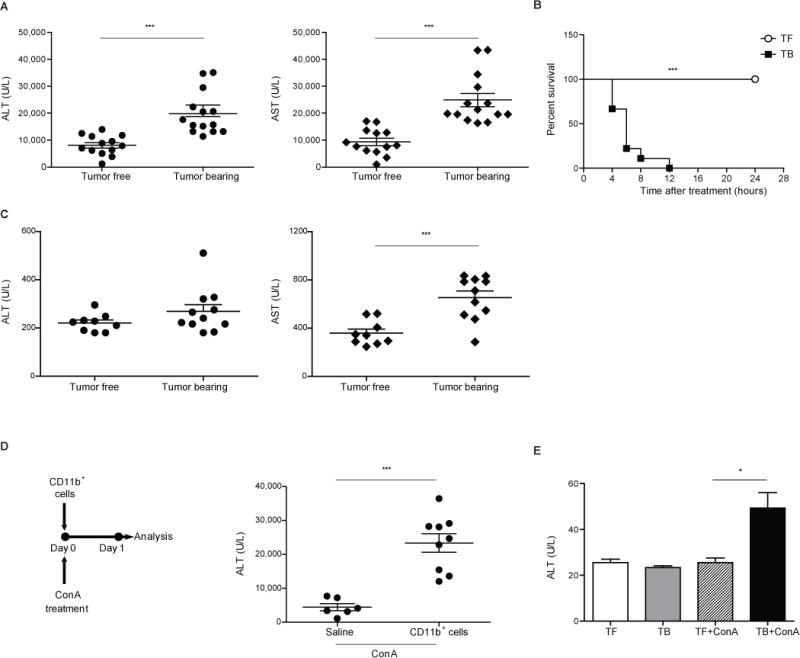
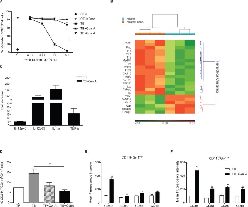
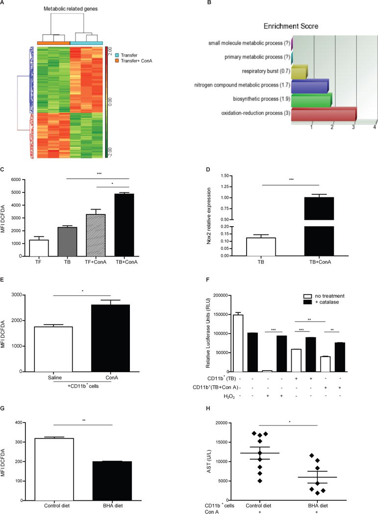
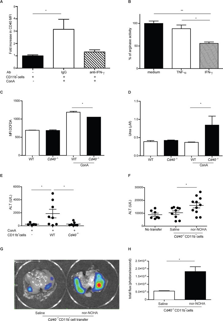
Immunosuppressive CD11b(+) Gr-1(+) myeloid-derived suppressor cells (MDSCs) accumulate in the livers of tumor-bearing (TB) mice. We studied hepatic MDSCs in two murine models of immune-mediated hepatitis. Unexpectedly, treatment of TB mice with Concanavalin A (Con A) or α-galactosylceramide resulted in increased alanine aminotransferase (ALT) and aspartate aminotransferase (AST) serum levels in comparison to tumor-free mice. Adoptive transfer of hepatic MDSCs into naïve mice exacerbated Con A induced liver damage. Hepatic CD11b(+) Gr-1(+) cells revealed a polarized proinflammatory gene signature after Con A treatment. An IFN-γ-dependent upregulation of CD40 on hepatic CD11b(+) Gr-1(+) cells along with an upregulation of CD80, CD86, and CD1d after Con A treatment was observed. Con A treatment resulted in a loss of suppressor function by tumor-induced CD11b(+) Gr-1(+) MDSCs as well as enhanced reactive oxygen species (ROS)-mediated hepatotoxicity. CD40 knockdown in hepatic MDSCs led to increased arginase activity upon Con A treatment and lower ALT/AST serum levels. Finally, blockade of arginase activity in Cd40(-/-) tumor-induced myeloid cells resulted in exacerbation of hepatitis and increased ROS production in vivo. Our findings indicate that in a setting of acute hepatitis, tumor-induced hepatic MDSCs act as proinflammatory immune effector cells capable of killing hepatocytes in a CD40-dependent manner.

Keywords: CD40; Concanavalin A; Immune-mediated hepatitis; Myeloid-derived suppressor cells; Reactive oxygen species; α-Galactosylceramide.


Figures

Figure 1
Figure 1. Tumor-bearing mice develop more severe immune-mediated hepatitis than tumor-free littermates
(A) Serum ALT/AST levels in TF (n=13) and EL4 TB (n=14) mice 16 hours after Con A treatment. (B) Kaplan-Meyer survival curve for cohorts of TF (n=5) and B16 GM-CSF TB mice (n=9) after Con A injection. (C) Serum ALT/AST levels in TF (n=7) and EL4 TB mice (n=8) 16 hours after α-GalCer injection. (D) Serum ALT values 16 hours after adoptive cell transfer of 5×10⁷ hepatic CD11b+ cells from B16 GM-CSF TB mice and Con A challenge (saline n=6; CD11b+ cells n=9). (E) TF (n=6) and EL4 TB (n=8) Rag-1−/− mice were injected either with saline or Con A. Serum ALT levels were determined as indicated above. Data are expressed as a mean ± SEM and are a cumulative of 6 (A), 2 (B), 3 (C–E) independent experiments. *P<0.05, ** P<0.01, ***P<0.001: Student’s t test (A,C, D and E) and Mantel-Cox log-rank test (B). TF = tumor-free; TB = tumor-bearing.
Figure 2
Figure 2. Immune-mediated hepatitis shapes MDSC into inflammatory myeloid cells
(A) Hepatic CD11b+Gr-1+ cells sorted from the livers of TF and EL4 TB mice, before or 3 hours after Con A challenge (n=4 mice/group) were cultured with 105 CFSE-labeled OT-I splenocytes at different ratios in the presence of OVA257–264 peptide (0.1 μg/ml). Proliferation of CFSE+CD8+ cells was evaluated after 48 hours. Data shown are mean ± SEM representative of 2 independent experiments. (B) Analysis of inflammation–related gene expression in EL4-induced CD45.1+ hepatic CD11b+Gr-1+ cells isolated from congenic mice 3 hours after adoptive transfer and induction of immune-mediated hepatitis. Dendrogram showing significant inflammation –related genes differentially expressed with fold change ≥ 2 Gene ontology score (n=7/group, pool of 3 independent experiments). Sample processing is described in Materials and Methods. (C) Increase in mRNA expression of several pro inflammatory genes in sorted liver CD11b+Gr-1+ cells from EL4 TB mice 3 hours after acute hepatitis induction (n=8 mice/ group; mRNA expression in sorted hepatic CD11b+Gr-1+ MDSC from TB mice without Con A injection was set to 1). Data shown are mean ± SEM representative of 2 independent experiments. (D) CD244 expression on hepatic CD11b+Gr-1high cells from either TF or TB mice before and 3 h after Con A challenge (n=2–4 mice/group). Cumulative data expressed as mean ± SEM are shown. (E–F) Mean Fluorescence Intensity (MFI) of CD40, CD80, CD86 and CD1d was determined either on CD11b+Gr-1high (E) or CD11b+Gr-1low (F) cells derived from EL4 TB mice, before or 3 hours after Con A treatment (n=3 mice/group). Data shown are mean ± SEM representative of 2 independent experiments *P<0.05, ** P<0.01, *** P<0.001: Student’s t test.
Figure 3
Figure 3. Liver CD11b+Gr-1+ MDSC produce hepatocyte damage via ROS release
(A) Dendrogram showing significant metabolism –related genes differentially expressed with fold change ≥ 2 Gene ontology score in metabolic processes (n=7/group, pool of 3 independent experiments). (B) Enrichment score bar chart grouping metabolism-related genes in metabolic processes. (C) ROS production was evaluated by flow cytometry gating on hepatic CD11b+Gr-1+ cells from TF and EL4 TB mice, before and 3 hours after Con A treatment (n=2–4 mice/group). (D) NADPH Oxidase 2 (Nox2) mRNA expression on EL4-induced hepatic CD11b+Gr-1+ cells before and 3 hours after Con A treatment (n=2–4 mice/group). (E) ROS production by flow cytometry gating on hepatic CD11b+Gr-1+ in adoptively transferred clonotypic B16 GM-CSF-induced hepatic CD11b+ cells with or without Con A co-injection. (F) Luminescence intensity in luciferase-expressing RIL-175 cells cultured with or without hepatic CD11b+ cells derived from EL4 TB mice before and 3 hours after Con A challenge (n=3 mice/group). 1000U/ml catalase was used to block ROS production and 2 mM H2O2 was set as positive control. (G–H) B16 GM-CSF TB mice were subjected for 3 days to a butylated hydroxyanisole (BHA) diet (7 mg/g bodyweight) or control diet (n=3 mice/group). ROS production gated on hepatic CD11b+Gr-1+ cells was evaluated by flow cytometry (G). Hepatic myeloid cells were MACS isolated (>70% are CD11b+Gr-1+ cells) and adoptively transferred followed by Con A challenge. AST levels were measured after 16 hours (H). Data are shown as mean ± SEM derived from representative (A–D, F–G) and cumulative (E,H) of 3 (A–C) and 2 (D–H) independent experiments. *P<0.05, ** P<0.01, *** P<0.001: Student’s t test.
Figure 4
Figure 4. Immune-mediated hepatitis changes hepatic MDSC into inflammatory myeloid cells by CD40 ligation
(A) CD40 MFI gated on CD45.1+CD11b+Gr-1+ cells 3 hours after congenic transfer of B16 GM-CSF-induced hepatic CD45.1+CD11b+ cells into CD45.2+ mice and Con A challenge. Data shown are mean ± SEM of 2 independent experiments. (B) Liver CD11b+ cells from B16 GM-CSF TB mice (n=3) were incubated for 16 hours in the presence of 0.1 μg/ml TNF-α or IFN-γ or were left untreated. Arginase activity was evaluated based on amount of urea release by the cell lysates. Data from untreated cells were set as 100%. Data shown are mean ± SEM representative of 2 independent experiments. (C–D) Function and phenotype of hepatic MDSC from either WT or Cd40−/− B16 GM-CSF TB mice before or 3 hours after Con A challenge (n=3/group): (C) ROS production ex vivo gated on hepatic CD11b+Gr-1+cells was evaluated by flow cytometry using Carboxy-H2-DCFDA. (D) Arginase activity was evaluated after overnight incubation of B16 GM-CSF TB liver CD11b+ cells. Data shown in C–D are mean ± SEM representative of at least 2 independent experiments. (E) 5×10⁷ liver CD11b+ cells from either WT or Cd40−/− B16 GM-CSF TB mice (n=3/group) were injected into naïve WT recipients and Con A was administered immediately. ALT (E) levels 16 hours after Con A are shown (n=5–8 mice/group). Cumulative data expressed as mean ± SEM, representative of 2 independent experiments. (F) liver CD11b+ cells from Cd40−/− B16 GM-CSF TB mice (n=3) were isolated and incubated either with PBS or arginase inhibitor nor-NOHA for 1 hour. Then 5×10⁷ cells were injected into naïve recipients followed by Con A injection. ALT levels 16 hours after Con A are shown (n=5–8 mice/group). Cumulative data expressed as mean ± SEM, representative of 2 independent experiments; (G–H) Mice treated as described in (F), were inoculated with 5 μg L-012, which shows luminescence after chemical reaction with ROS. Data show a representative in vivo image (G) and quantification of bioluminescence (H) (n=3 mice/group). Cumulative data expressed as mean ± SEM, representative 2 independent experiments. *P<0.05, *** P<0.001: Student’s t test.

Similar articles

Cited by

References

    1. Talmadge JE, Gabrilovich D. History of myeloid-derived suppressor cells. Nat Rev Cancer. 2013;13:7752. doi: 10.1038/nrc3581. - DOI - PMC - PubMed
    1. Gabrilovich DI, Bronte V, Chen S-H, Colombo MP, Ochoa A, Ostrand-Rosenberg S, Schreiber H. The terminology issue for myeloid-derived suppressor cells. Cancer Res. 2007;67:425. doi: 10.1158/0008-5472.CAN-06-3037. author reply 426. - DOI - PMC - PubMed
    1. Tiegs G, Hentschel J, Wendel A. A T cell-dependent experimental liver injury in mice inducible by concanavalin A. Journal of Clinical Investigation. 1992;90:196–203. doi: 10.1172/JCI115836. - DOI - PMC - PubMed
    1. Mizuhara H, O’Neill E, Seki N, Ogawa T, Kusunoki C, Otsuka K, Satoh S, et al. T cell activation-associated hepatic injury: mediation by tumor necrosis factors and protection by interleukin 6. J Exp Med. 1994;179:1529–1537. - PMC - PubMed
    1. Gabrilovich DI, Ostrand-Rosenberg S, Bronte V. Coordinated regulation of myeloid cells by tumours. Nat Rev Immunol. 2012;12:253–268. doi: 10.1038/nri3175. - DOI - PMC - PubMed

Publication types

MeSH terms