Review
. 2015 Jul:54:89-107.
doi: 10.1016/j.neubiorev.2015.01.014. Epub 2015 Jan 20.

Limbic circuitry of the midline thalamus

Affiliations
Review

Limbic circuitry of the midline thalamus

Robert P Vertes et al. Neurosci Biobehav Rev. 2015 Jul.

Abstract

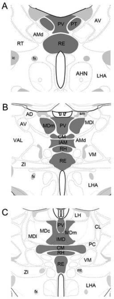
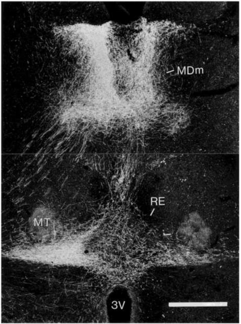
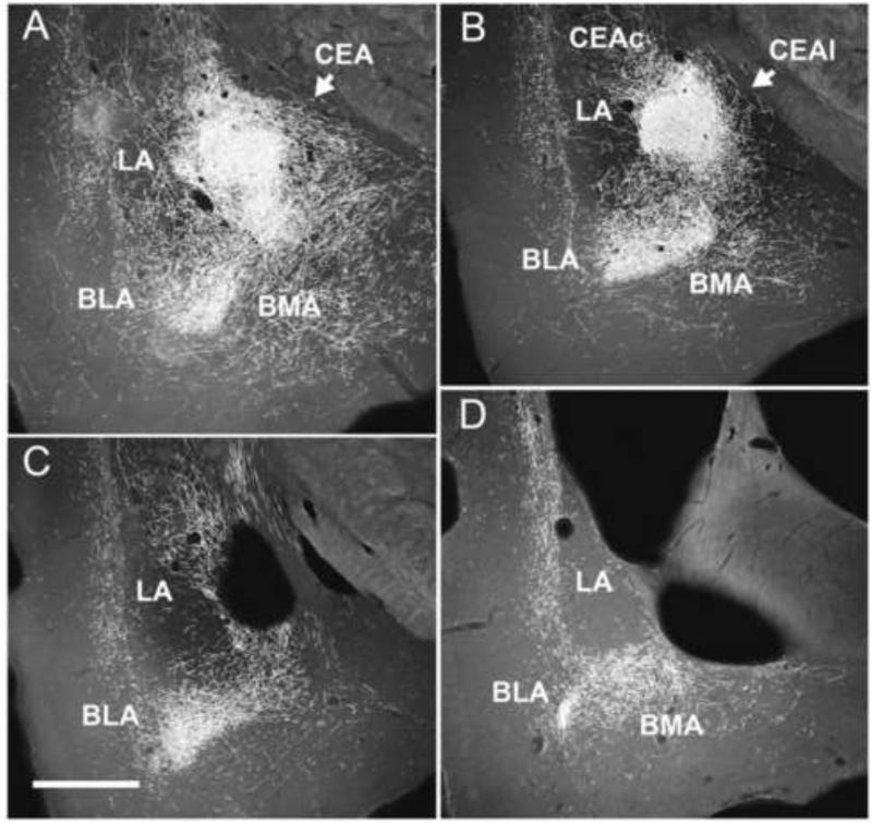
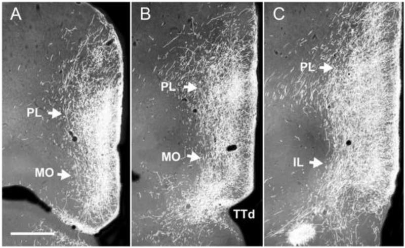
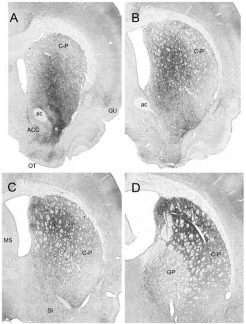
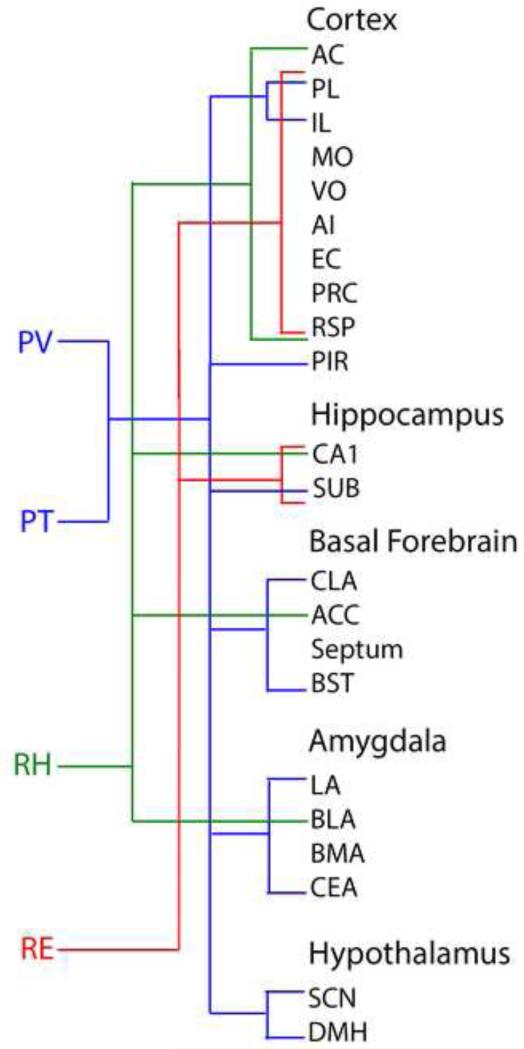
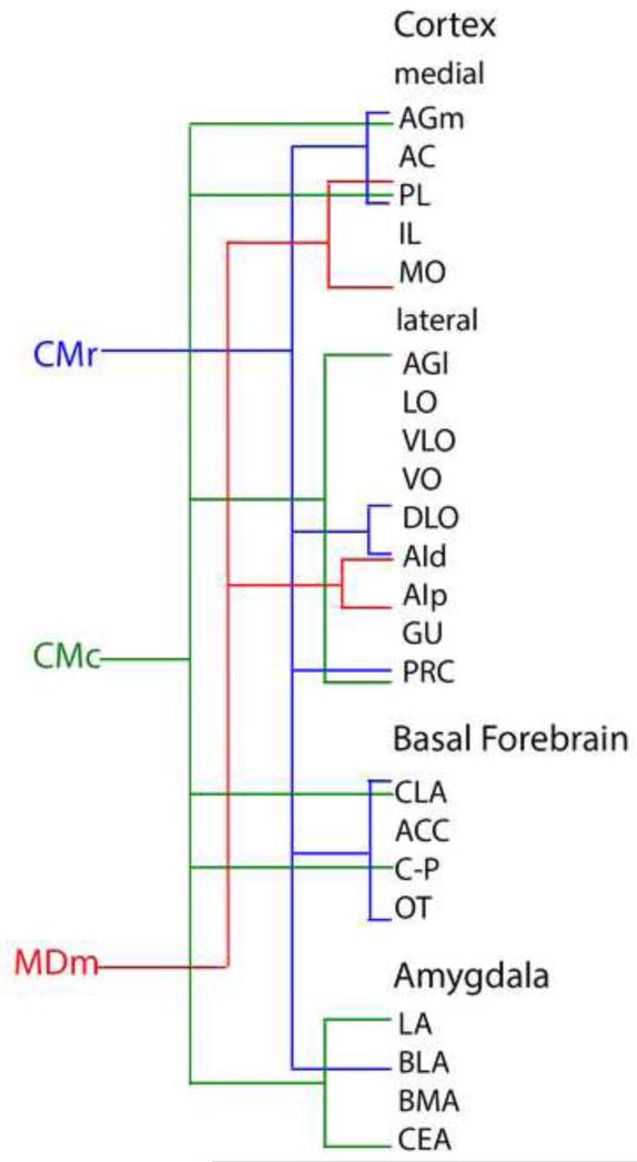
The thalamus was subdivided into three major groups: sensorimotor nuclei (or principal/relay nuclei), limbic nuclei and nuclei bridging these two domains. Limbic nuclei of thalamus (or 'limbic thalamus') consist of the anterior nuclei, midline nuclei, medial division of the mediodorsal nucleus (MDm) and central medial nucleus (CM) of the intralaminar complex. The midline nuclei include the paraventricular (PV) and paratenial (PT) nuclei, dorsally, and the reuniens (RE) and rhomboid (RH) nuclei, ventrally. The 'limbic' thalamic nuclei predominantly connect with limbic-related structures and serve a direct role in limbic-associated functions. Regarding the midline nuclei, RE/RH mainly target limbic cortical structures, particularly the hippocampus and the medial prefrontal cortex. Accordingly, RE/RH participate in functions involving interactions of the HF and mPFC. By contrast, PV/PT mainly project to limbic subcortical structures, particularly the amygdala and nucleus accumbens, and hence are critically involved in affective behaviors such as stress/anxiety, feeding behavior, and drug seeking activities. The anatomical/functional characteristics of MDm and CM are very similar to those of the midline nuclei and hence the collection of nuclei extending dorsoventrally along the midline/paramidline of the thalamus constitute the core of the 'limbic thalamus'.

Keywords: Affect; Central medial nucleus; Cognition; Drug seeking activity; Feeding behavior; Learning and memory; Mediodorsal nucleus; Nucleus reuniens; Paratenial nucleus; Paraventricular nucleus; Rhomboid nucleus; Stress/anxiety.


Figures

Figure 1
Figure 1
Schematic representation of three rostrocaudally (A-C) aligned sections through the diencephalon depicting (in dark gray) the locations of dorsoventrally oriented nuclei along the midline/paramidline of the thalamus, which together with the anterior nuclei of thalamus, constitute the ‘limbic thalamus’. They include the paraventricular (PV), paratenial (PT), reuniens (RE) and rhomboid (RH) nuclei of the midline thalamus, the medial division of the mediodorsal nucleus (MDm), the central medial nucleus (CM) of the rostral intralaminar complex, the interanteromedial nucleus (IAM) and the intermediodorsal (IMD) nucleus. Sections modified from Swanson (2004). See list for abbreviations.
Figure 2
Figure 2
Low magnification darkfield photomicrographs showing patterns of labeling in the dorsal (A) and ventral hippocampus (B,C) produced by an injection of PHA-L in nucleus reuniens of the midline thalamus. Note the dense concentration of labeled fibers restricted to the stratum lacunosum moleculare of CA1 of the dorsal (A) and ventral (B) hippocampus and the molecular layer of the ventral subiculum (C). See list for abbreviations. Scale bar for A = 600 μm; for B,C = 1000 μm. Modified from Vertes et al. (2006).
Figure 3
Figure 3
A,B: low magnification darkfield photomicrographs of transverse sections through the rostral forebrain showing patterns of labeling within the medial prefrontal cortex produced by an injection of PHA-L in nucleus reuniens of the midline thalamus. Note the presence of pronounced numbers of labeled fibers in the infralimbic (IL), prelimbic (PL), anterior cingulate (AC) and medial (frontal) agranular (AGm) cortices, stronger in IL and PL than in AC and AGm Note additional labeling in the claustrum (CLA), the rostral pole of nucleus accumbens (ACC) and the dorsal agranular insular cortex (AId). C: high magnification darkfield photomicrograph from A (arrows) showing labeled fibers in all layers of IL, PL and AC, most densely concentrated in layers 1 and 5/6, and mediolaterally aligned fibers in the middle layers of these fields, oriented parallel to the cell layers. See list for abbreviations. Scale bar for A,B = 1000 μm; for C = 250 μm. Modified from Vertes et al. (2006).
Figure 4
Figure 4
Low (A) and high (B,C) magnification light-field photomicrographs depicting retrogradely labeled cells in the ventral subicular complex of the hippocampus produced by a Fluorogold injection in nucleus reuniens of the midline thalamus. As shown, pronounced numbers of labeled cells extended dorsal-ventrally throughout the subiculum (SUB) within the ventral subiculum and the postsubiculum (POST). B,C: Clusters of labeled cells dorsally in the ventral subiculum (arrow in A) at higher levels of magnification. See list for abbreviations. Scale bar for A = 325 μm; for B,C = 65 μm. Modified from McKenna and Vertes (2004).
Figure 5
Figure 5
Darkfield photomicrograph of a transverse section through the diencephalon showing patterns of labeling within nuclei of the midline thalamus produced by a PHA-L injection in the infralimbic cortex. Note dense labeling virtually confined to the paraventricular and medial division of the mediodorsal nucleus (MDm), dorsally, and the nucleus reuniens (RE), ventrally. See list for abbreviations. Scale bar = 450 μm. Modified from Vertes (2002).
Figure 6
Figure 6
Series of electron micrographic sections through the nucleus reuniens of the midline thalamus (a-d) showing asymmetric contacts of a single PHA-L labeled (F) fibers from the medial prefrontal cortex onto a labeled dendritic shaft, identified by the presence of numerous silver intensified gold deposits (arrows in D) of a nucleus reuniens cell retrogradely from a Fluorogold injection in the ventral hippocampus. Note also the presence of asymmetric contacts of an unlabeled fiber (A) on the same labeled dendrite segment (D). Scale bar, 1 μm. Modified from Vertes et al. (2007).
Figure 7
Figure 7
Low (A) and high (B) magnification darkfield photomicrographs of a transverse section through the anterior forebrain showing patterns of labeling in the dorsal and ventral striatum produced by a PHA-L injection in the rhomboid nucleus of the midline thalamus. B taken from A (arrowheads). Note dense labeling in the nucleus accumbens (ACC) and adjacent ventromedial parts of the dorsal striatum. See list for abbreviations. Scale bar: for A = 750 μm; for B = 400 μm. Modified from Vertes et al. (2006).
Figure 8
Figure 8
Low magnification darkfield photomicrographs of transverse sections through anterior forebrain showing patterns of labeling in the nucleus accumbens (ACC) (A) and the bed nucleus of the stria terminalis (BST) (B) produced by a PHA-L injection in the anterior paraventricular nucleus of the midline thalamus. Note the pronounced labeling in both the shell and core of ACC, with additional labeling in the adjacent lateral septum (LS) as well as in BST above and below the anterior commissure. See list for abbreviations. Scale bar = 500 μm. Modified from Vertes and Hoover (2008).
Figure 9
Figure 9
A-D: Series of low magnification darkfield photomicrographs of transverse sections rostrocaudally (A-D) through the forebrain depicting patterns of labeling within the amygdala produced by a PHA-L injection in the posterior paraventricular nucleus of the midline thalamus. A,B: Note dense labeling in the central (CEA), basomedial (BMA) and basolateral (BLA) nuclei of amygdala, and marked but less pronounced labeling in parts of the medial, lateral (LA) and anterior cortical nuclei of the amygdala. C,D: Note prominent labeling caudally within the amygdala mainly confined to BMA and BLA. See list for abbreviations. Scale bar = 750 μm. Modified from Vertes and Hoover (2008).
Figure 10
Figure 10
A-C: Series of rostrocaudally (A-C) aligned low magnification darkfield photomicrographs of transverse sections through the anterior forebrain depicting patterns of labeling within the medial prefrontal cortex (mPFC) produced by a PHA-L injection into the paratenial nucleus of the midline thalamus. Note intense labeling along the ventral medial wall of the mPFC mainly confined to the prelimbic (PL) (A-C), medial orbital (MO) (A,B) and infralimbic (C) (IL) cortices. As depicted, labeling was particularly dense in layers 1 and 3 of these prefrontal cortical fields. See list for abbreviations. Scale bar = 750 μm. Modified from Vertes and Hoover (2008).
Figure 11
Figure 11
Low power lightfield photomicrographs of four rostrocaudally aligned (A-D) transverse sections through the rostral forebrain showing patterns of labeling in the dorsal striatum (C-P) produced by a PHA-L injection rostrally in the central medial nucleus of the thalamus. Note dense labeling spread throughout C-P, concentrated rostrally (A,B), dorsoventrally within the mid-central C-P, and caudally (C,D) within the medial/dorsomedial half of C-P. See list for abbreviations. Scale bar for A = 975 μm; for B,C = 1000 μm; for D = 950 μm. Modified from Vertes et al. (2012).
Figure 12
Figure 12
Summary diagram comparing and contrasting the main projection sites of the paraventricular PVP, paratenial (PT), reuniens (RE) and rhomboid (RH) nuclei of the midline thalamus to the cortex, basal forebrain, amygdala and hypothalamus. See list for abbreviations.
Figure 13
Figure 13
Summary diagram comparing and contrasting the main projection sites of the rostral (CMr) and caudal (CMc) central medial nucleus and the medial division of the mediodorsal nucleus (MDm) of thalamus to the cortex, basal forebrain and amygdala. See list for abbreviations.

References

    1. Aggleton JP, Dumont JR, Warburton EC. Unraveling the contributions of the diencephalon to recognition memory: a review. Learn. Mem. 2011;18:384–400. - PMC - PubMed
    1. Angeles-Castellanos M, Mendoza J, Escobar C. Restricted feeding schedules phase shift daily rhythms of c-Fos and protein Per1 immunoreactivity in corticolimbic regions in rats. Neuroscience. 2007;144:344–355. - PubMed
    1. Bailey KR, Mair RG. Lesions of specific and nonspecific thalamic nuclei affect prefrontal cortex-dependent aspects of spatial working memory. Behav. Neurosci. 2005;119:410–419. - PubMed
    1. Barker GR, Warburton EC. When is the hippocampus involved in recognition memory? J. Neurosci. 2011;31:10721–10731. - PMC - PubMed
    1. Barker GR, Bird F, Alexander V, Warburton EC. Recognition memory for objects, place, and temporal order: a disconnection analysis of the role of the medial prefrontal cortex and perirhinal cortex. J. Neurosci. 2007;27:2948–2957. - PMC - PubMed

Publication types