. 2015 Mar;14(3):297-310.
doi: 10.1128/EC.00238-14. Epub 2015 Jan 23.

The ADP/ATP carrier and its relationship to oxidative phosphorylation in ancestral protist trypanosoma brucei

Affiliations

The ADP/ATP carrier and its relationship to oxidative phosphorylation in ancestral protist trypanosoma brucei

Anna Gnipová et al. Eukaryot Cell. 2015 Mar.

Abstract

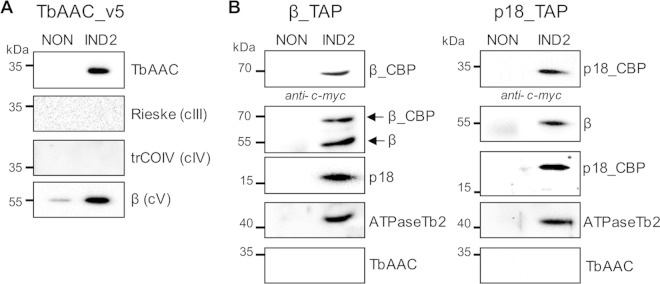
The highly conserved ADP/ATP carrier (AAC) is a key energetic link between the mitochondrial (mt) and cytosolic compartments of all aerobic eukaryotic cells, as it exchanges the ATP generated inside the organelle for the cytosolic ADP. Trypanosoma brucei, a parasitic protist of medical and veterinary importance, possesses a single functional AAC protein (TbAAC) that is related to the human and yeast ADP/ATP carriers. However, unlike previous studies performed with these model organisms, this study showed that TbAAC is most likely not a stable component of either the respiratory supercomplex III+IV or the ATP synthasome but rather functions as a physically separate entity in this highly diverged eukaryote. Therefore, TbAAC RNA interference (RNAi) ablation in the insect stage of T. brucei does not impair the activity or arrangement of the respiratory chain complexes. Nevertheless, RNAi silencing of TbAAC caused a severe growth defect that coincides with a significant reduction of mt ATP synthesis by both substrate and oxidative phosphorylation. Furthermore, TbAAC downregulation resulted in a decreased level of cytosolic ATP, a higher mt membrane potential, an elevated amount of reactive oxygen species, and a reduced consumption of oxygen in the mitochondria. Interestingly, while TbAAC has previously been demonstrated to serve as the sole ADP/ATP carrier for ADP influx into the mitochondria, our data suggest that a second carrier for ATP influx may be present and active in the T. brucei mitochondrion. Overall, this study provides more insight into the delicate balance of the functional relationship between TbAAC and the oxidative phosphorylation (OXPHOS) pathway in an early diverged eukaryote.


Figures

FIG 1
FIG 1
TbAAC colocalizes with higher-molecular-weight complexes. (A) The hypotonically purified mitochondria were solubilized using digitonin (0.5 mg, 1 mg, and 2 mg detergent/mg protein) and separated on a 3 to 8% hrCN PAGE. Isolated lanes from these native gels were further processed in the second dimension by denaturing Tricine–SDS-PAGE, and the gel was blotted on a nitrocellulose membrane for immunodetection using a polyclonal anti-TbAAC antibody. “Front” indicates the leading edge of the first-dimensional gel. The sizes of the native molecular mass markers are indicated on top of the first panel. The protein marker for the second dimension is indicated on the left. (B) The nitrocellulose membrane containing mitochondria lysed with 1 mg digitonin/mg mt protein was stripped and further probed individually with trypanosome-specific polyclonal antibodies against subunit COVI (complex IV), subunit β (FoF1-ATP synthase), and PiC. The positions of monomeric and oligomeric FoF1-ATP synthase and complex IV are denoted by arrows. The resulting compilation of Western images was overlaid using Adobe Photoshop.
FIG 2
FIG 2
TbAAC does not copurify with complex IV or FoF1-ATP synthase. (A) TbAAC_v5-tagged complexes were purified using magnetic beads (Dynabeads M-280, sheep anti-mouse IgG) charged with a monoclonal anti-v5 antibody. The tagged protein complexes were purified from noninduced (NON) cells and 2-day Tet-induced cells (IND2) containing the regulatable ectopic v5-tagged TbAAC protein. The tagged protein complexes were eluted with SDS-PAGE loading buffer, fractionated by SDS-PAGE, and examined by Western blotting. The presence of the tagged TbAAC_v5 was verified using an anti-TbAAC antibody (top). The association of TbAAC_v5 with core subunits of complex III (Rieske), IV (trCOIV), and FoF1-ATPase (β) was determined using specific antibodies. The applicable sizes of the protein marker are indicated on the left. (B) FoF1-ATP synthase complexes were purified using tandem affinity purification from noninduced (NON) cells and cells induced for 2 days (IND2) to express the ectopically tagged subunits β_TAP (left) and p18_TAP (right). Tagged FoF1-ATP synthase complexes were purified by IgG affinity chromatography, eluted by TEV protease, resolved by SDS-PAGE, and examined by Western blotting. The presence of cleaved bait proteins now containing just the calmodulin-binding protein (CBP) and c-myc epitope tag, β_CBP and p18_CBP, was verified by an anti-c-myc antibody. Blots probed with antibodies specific to subunits β, p18, and ATPaseTb2 demonstrate that the TEV eluates contain components of both the F1 and Fo moieties of the FoF1-ATP synthase (endogenous and TAP tagged), while Western blots immunodecorated with an anti-TbAAC antibody demonstrate that the ADP/ATP transporter is not detectable in β- and p18-tagged complexes. Noninduced cells were used as a negative control for any nonspecific protein binding during the IgG affinity purification. The relevant sizes of the protein marker are indicated on the left.
FIG 3
FIG 3
TbAAC is essential for in vitro growth but does not impair either the steady-state abundance or sedimentation profile of OXPHOS complexes or the in-gel activities of complex IV and FoF1-ATPase. (A) Growth curves of the noninduced (NON) and Tet-induced (IND) TbAAC RNAi procyclic T. brucei cell lines. Cells were maintained in the exponential growth phase (between 106 and 107 cells/ml), and cumulative cell numbers were calculated from daily counts that incorporated each subsequent dilution factor. The inset depicts a Northern blot analysis of TbAAC mRNA levels in the parental 29-13 cells, noninduced cells (NON), and cells induced for 2 days of RNAi (IND2). (B) The steady-state abundance of TbAAC, core subunits of complex III (apoC and Rieske), complex IV (trCOIV and COVI), FoF1-ATP synthase (β), and TbPiC in noninduced (NON) TbAAC RNAi cells and those induced for 1, 3, and 5 days (IND1, IND3, and IND5) was determined by Western blotting using specific polyclonal antibodies. The numbers beneath the TbAAC blot represent the abundance of immunodetected TbAAC, expressed as a percentage of the noninduced samples after normalization to the loading control, mt Hsp70. The relevant sizes of the protein marker are on the left. The TbPiC antiserum cross-reacts with TbAAC, and this band is indicated with an asterisk. (C) The sedimentation profiles of TbAAC, TbPiC, complexes III and IV, and FoF1-ATP synthase were examined using Western blot analysis of glycerol gradient fractions. Mitochondria from non-RNAi-induced cells (NON) and cells induced for 5 days (IND5) were lysed with dodecyl maltoside. An equal amount of the cleared lysate from 1 × 10⁹ trypanosomes was loaded on each 10-to-30% glycerol gradient. Western analyses with trypanosomatid-specific antibodies raised against AAC, PiC, apoC (complex III), trCOIV (complex IV), and subunit β (FoF1-ATP synthase) depict the sedimentation profile of the examined OXPHOS complexes. The glycerol gradient fractions are numbered, and the sizes of the protein marker are indicated. An asterisk designates the TbAAC band that cross-reacts with the TbPiC antiserum. (D) In-gel complex IV and FoF1-ATP synthase activity staining after TbAAC repression. mt preparations from noninduced (NON) and RNAi cells Tet-induced for 5 days (IND5) were solubilized using dodecyl maltoside and separated either by 6% BN PAGE for complex IV staining or by 3-to-12% BN PAGE for FoF1-ATPase staining. The arrows denote bands visualized by the specific activity staining. The sizes of high-molecular-weight markers (ferritin and its dimer; Sigma) are on the left (in kilodaltons).
FIG 4
FIG 4
Both ADP and ATP influx into the mitochondrion are affected in cells silenced for TbAAC expression. (A) To determine levels of ADP influx into the mitochondrion, the in vitro production of ATP was measured in digitonin-extracted mitochondria from noninduced (NON) cells or cells with activated TbAAC RNAi for 3 (IND3) or 5 days (IND5). The phosphorylation pathways were triggered by the addition of ADP and one of the following substrates: succinate, α-ketoglutarate, and pyruvate/succinate. Malonate (MAL), a specific inhibitor of succinate dehydrogenase, was used to inhibit ATP production by oxidative phosphorylation, and atractyloside (ATR) was used to inhibit import of ADP into mitochondria. The level of ATP production in mitochondria isolated from noninduced RNAi cells stimulated with substrate, but in the absence of any specific inhibitors, was established as the reference and set to 100%. All other values for the same substrate are calculated arithmetic means expressed as percentages of this reference sample. The “No ADP” value serves as a control for the background production of ATP from endogenous mt ADP. The data are averages from at least three independent experiments and standard deviations. (B) To ascertain the extent of ATP import into active mitochondria, ATPase hydrolytic activities were measured in either noninduced (NON) TbAAC RNAi cells or cells induced with Tet for 3 (IND3) and 5 days (IND5). Crude mt vesicles were obtained by digitonin extraction. The ATPase activity was triggered by ATP addition (5 mM) and assayed by measuring the release of free phosphate. The total amount of free phosphate created from all ATPase enzymes present in the noninduced sample was set at 100%. The displayed results represent the average activities obtained from extracts prepared from four independent RNAi inductions. (C) Total ATPase activity measured in mitochondrial lysates from noninduced and RNAi cells induced for 5 days indicate that mt ATPases are still active when substrate is available. The total amount of free phosphate created from all ATPase enzymes present in the noninduced sample was set at 100%. The results are the average activities obtained from extracts prepared from four independent RNAi inductions.
FIG 5
FIG 5
Cytosolic ATP levels are significantly decreased in TbAAC RNAi-induced cells. (A) The subcellular localization of luciferase was determined within TbAAC RNAi cells expressing luciferase (LUC-TbAAC RNAi). Parental TbAAC RNAi cells were used as a negative control. T. brucei cells (5 × 10⁸) were harvested by centrifugation (WCL) and further separated into cytosolic (C) and mitochondrial (M) subcellular fractions upon digitonin extraction. Purified fractions were analyzed by Western blotting with anti-luciferase, anti-mt Hsp70 (mitochondria), and anti-enolase (cytosol) antibodies. The relevant sizes of the protein markers are on the left. (B) Total cytosolic ATP content was measured by a luminometer upon luciferin addition to living cells, either noninduced (NON) LUC-TbAAC RNAi cells or cells induced for 3 days (IND3). Values are in relative light units (RLU) and are averages from three independent RNAi inductions.
FIG 6
FIG 6
TbAAC silencing increases the Δψm, resulting in decreased oxygen consumption and elevated ROS production. (A) After staining with 60 nM TMRE, the Δψm was measured by flow cytometry in both noninduced cells (NON) and TbAAC RNAi cells induced for 3 (IND3), 5 (IND5), and 7 (IND7) days. The median fluorescent intensity values are presented as percentages of the value for the noninduced sample, which was set to 100%. Data were obtained from at least three independent RNAi experiments, and the standard deviations are included. (B) The ROS detection reagent DCFH-DA was quantified by fluorescence-activated cell sorting (FACS) analysis in noninduced (NON) cells and TbAAC RNAi cells induced for 3 (IND3), 5 (IND5), and 7 (IND7) days. Increased fluorescence intensity corresponds to an accumulated ROS formation that was observed in three independent assays. (C) The oxygen consumption of noninduced (NON) and RNAi-induced (IND5) cells incubated in SDM-79 medium at 27°C was monitored with a Clark-type oxygen electrode. The total oxygen consumption in noninduced cells was set to 100%. Values are arithmetic means and standard deviations from three experiments. (D) The ratio of complex IV- and TAO-mediated respiration was measured in noninduced (NON) and TbAAC RNAi cells induced for 5 days (IND5). The total oxygen consumption in both cell lines was set to 100%. After the addition of salicylhydroxamic acid (SHAM) (0.03 mM), a potent TAO inhibitor, the remaining oxygen consumption represented respiration occurring via complex IV. Values are means and standard deviations from six independent experiments.
FIG 7
FIG 7
TbAAC depletion leads to defects in cell division. (A) DAPI staining was used to visualize various nucleus/kinetoplast (NK) phenotypes in noninduced TbAAC RNAi cells and cells induced with Tet for 5 days. (Top) Normal cell types either in G1/S (1N1K) and G2/M (1N2K) phases or undergoing cytokinesis (2N2K). (Bottom) Representative abnormal cell types (1K, 2N1K, and xNxK). N, nucleus; K, kinetoplast. (B) Quantification of the microscopy images based on the number of nuclei and kinetoplasts in more than 200 cells per time point. NON, noninduced cells; IND5, RNAi cells induced for 5 days.

References

    1. Arco AD, Satrustegui J. 2005. New mitochondrial carriers: an overview. Cell Mol Life Sci 62:2204–2227. doi:10.1007/s00018-005-5197-x. - DOI - PMC - PubMed
    1. Klingenberg M. 2008. The ADP and ATP transport in mitochondria and its carrier. Biochim Biophys Acta 1778:1978–2021. doi:10.1016/j.bbamem.2008.04.011. - DOI - PubMed
    1. Dudkina NV, Kouril R, Peters K, Braun HP, Boekema EJ. 2010. Structure and function of mitochondrial supercomplexes. Biochim Biophys Acta 1797:664–670. doi:10.1016/j.bbabio.2009.12.013. - DOI - PubMed
    1. Dudkina NV, Heinemeyer J, Sunderhaus S, Boekema EJ, Braun HP. 2006. Respiratory chain supercomplexes in the plant mitochondrial membrane. Trends Plant Sci 11:232–240. doi:10.1016/j.tplants.2006.03.007. - DOI - PubMed
    1. Minauro-Sanmiguel F, Wilkens S, Garcia JJ. 2005. Structure of dimeric mitochondrial ATP synthase: novel F0 bridging features and the structural basis of mitochondrial cristae biogenesis. Proc Natl Acad Sci U S A 102:12356–12358. doi:10.1073/pnas.0503893102. - DOI - PMC - PubMed

Publication types

MeSH terms

Substances

LinkOut - more resources