. 2015 Feb 10;112(6):E556-65.
doi: 10.1073/pnas.1412058112. Epub 2015 Jan 23.

Effector Vγ9Vδ2 T cells dominate the human fetal γδ T-cell repertoire

Affiliations

Effector Vγ9Vδ2 T cells dominate the human fetal γδ T-cell repertoire

Tanya Dimova et al. Proc Natl Acad Sci U S A. .

Abstract

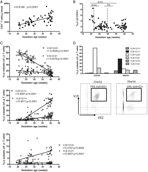
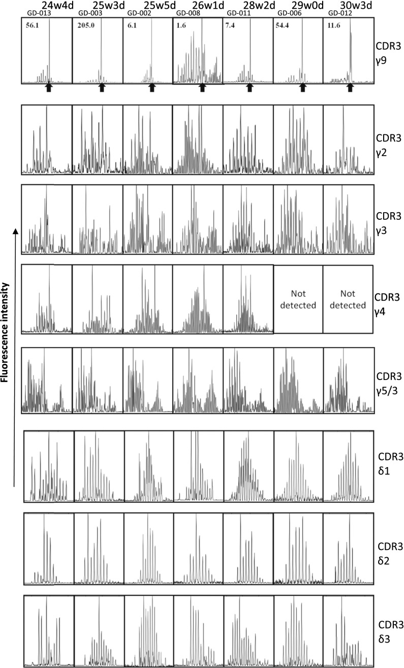
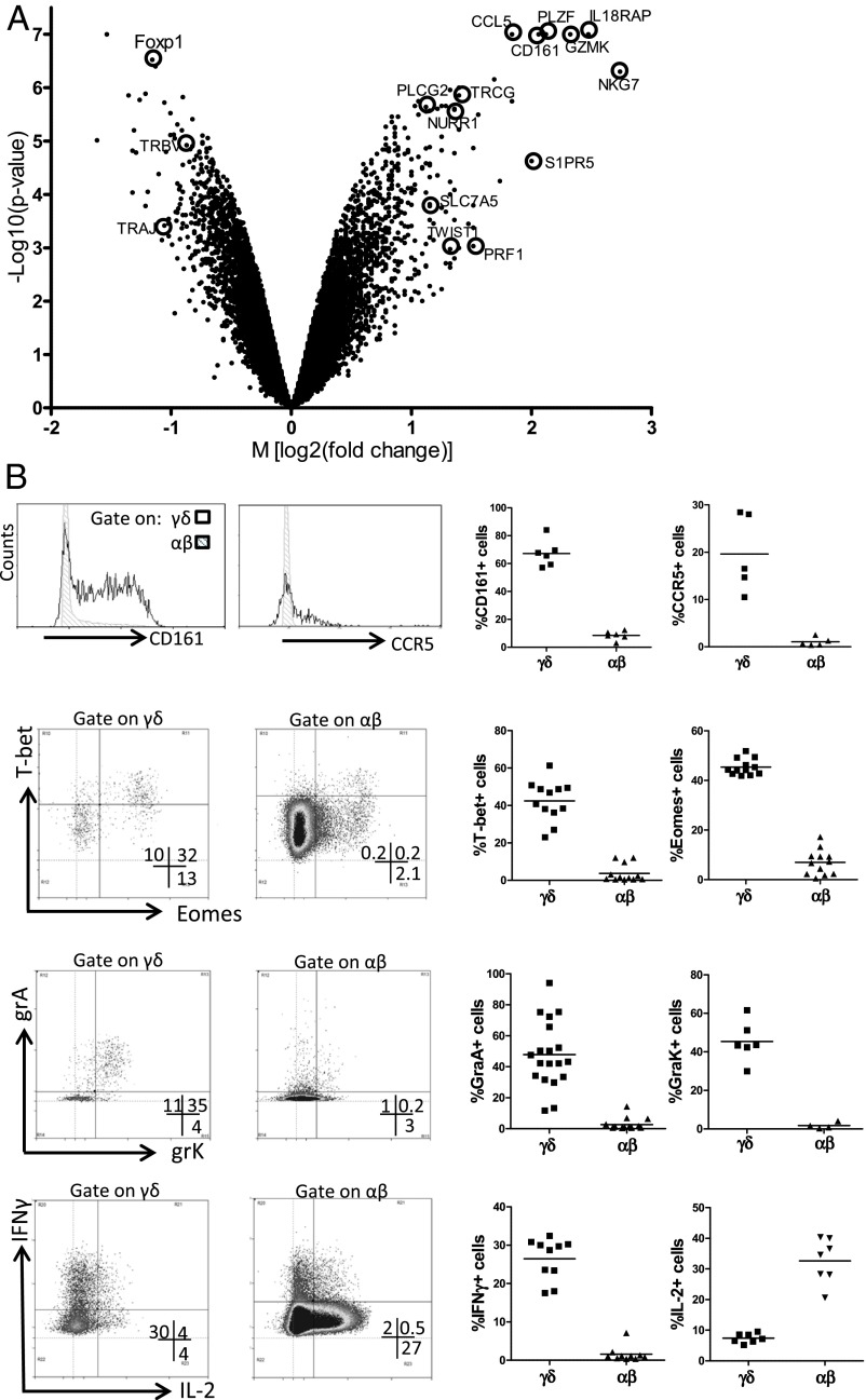
γδ T cells are unconventional T cells recognizing antigens via their γδ T-cell receptor (TCR) in a way that is fundamentally different from conventional αβ T cells. γδ T cells usually are divided into subsets according the type of Vγ and/or Vδ chain they express in their TCR. T cells expressing the TCR containing the γ-chain variable region 9 and the δ-chain variable region 2 (Vγ9Vδ2 T cells) are the predominant γδ T-cell subset in human adult peripheral blood. The current thought is that this predominance is the result of the postnatal expansion of cells expressing particular complementary-determining region 3 (CDR3) in response to encounters with microbes, especially those generating phosphoantigens derived from the 2-C-methyl-d-erythritol 4-phosphate pathway of isoprenoid synthesis. However, here we show that, rather than requiring postnatal microbial exposure, Vγ9Vδ2 T cells are the predominant blood subset in the second-trimester fetus, whereas Vδ1(+) and Vδ3(+) γδ T cells are present only at low frequencies at this gestational time. Fetal blood Vγ9Vδ2 T cells are phosphoantigen responsive and display very limited diversity in the CDR3 of the Vγ9 chain gene, where a germline-encoded sequence accounts for >50% of all sequences, in association with a prototypic CDR3δ2. Furthermore, these fetal blood Vγ9Vδ2 T cells are functionally preprogrammed (e.g., IFN-γ and granzymes-A/K), with properties of rapidly activatable innatelike T cells. Thus, enrichment for phosphoantigen-responsive effector T cells has occurred within the fetus before postnatal microbial exposure. These various characteristics have been linked in the mouse to the action of selecting elements and would establish a much stronger parallel between human and murine γδ T cells than is usually articulated.

Keywords: Vγ9Vδ2; fetus; gammadelta; human; neonate.


Conflict of interest statement

The authors declare no conflict of interest.

Figures

Fig. 1.
Fig. 1.
Human fetal peripheral blood is highly enriched for the presence of Vγ9Vδ2 T cells around 20 wk gestation. (A) Absolute numbers of T cells per microliter of blood as determined by flow cytometry on fresh blood using Trucount tubes (n = 64); the linear regression line is shown together with its r and P values. (B) Percentage of γδ T cells (of CD3+ lymphocytes) according to gestational age; total n = 87; comparisons are shown between 19w2d–21w2d (n = 7), 21w5d–29w5d (n = 40), and 30w3d–41w1d (n = 40). ***P < 0.001; ns, not significant. (C) Percentages of γδ T-cell subsets of γδ+CD3+ lymphocytes according to gestational age; linear regression lines are shown with their corresponding r and P values. (D) Flow cytometry data on γδ subset percentages of γδ+CD3+ lymphocytes for one fetus from which we obtained blood both at 23w5d and at 39w5d gestation; these data are representative fo the data for three different fetuses from which blood samples were derived at two different gestational times; in the lower panel, the gate is put on γδ+CD3+ lymphocytes.
Fig. 2.
Fig. 2.
The CDR3γ9 repertoire of fetal blood before 30 wk gestation is highly restricted and enriched for a 14-aa length. Each box represents the spectratyping data of one fetus of the indicated CDR3 chain. The fetuses (30 wk gestation or lower) are ordered from left to right by low to high gestational age. Within the CDR3γ9 spectratyping results, the ratio of Vγ9+Vδ2+/Vγ9+Vδ2 cells is indicated for each fetus at the left top corner of the box; arrows indicate the highly enriched length of 14 aa.
Fig. 3.
Fig. 3.
Fetal Vγ9Vδ2 T cells specifically possess a highly restricted germline-encoded CDR3γ9 using JγP and a conserved highly hydrophobic residue at position 5 of their CDR3δ2. (A) A typical flow cytometry plot of Vγ9 vs. Vδ2 staining on fetal blood γδ T cells (before 30 wk gestation) with gate settings illustrating the sorting strategy to sort Vγ9+Vδ2+ γδ T cells and nonVγ9Vδ2 γδ T cells; the gate is preset on CD3+γδ+ cells. (B) CDR3γ9 (Left) and CDR3δ2 (Right) showing the spectratyping plots of the sorted subsets of three different fetuses. (C) Comparison of sequence features of CDR3γ9 and CDR3δ2 in Vγ9Vδ2 versus non-Vγ9Vδ2 subsets of three fetuses.
Fig. 4.
Fig. 4.
In vitro exposure to endogenous and exogenous phosphoantigens leads to the expansion of fetal Vγ9Vδ2 T cells expressing the public/invariant CDR3γ9 CALWEVQELGKKIKVF. (A and B) Exposure of fetal PBMC to zoledronate (10 μM) in the presence of 100 U/mL IL-2 (A) and HMB-PP (100 μM) in the presence of 100 U/mL IL-2 and 50 ng/mL IL-18 (B, Upper) for 10 d. Flow cytometric staining for Vγ9 and Vδ2 ex vivo (Left) and after in vitro culture (Right); the gate is preset on CD3+ lymphocytes; numbers indicate the percentage of CD3+ lymphocytes that are Vγ9+Vδ2+. Similar results were obtained counting absolute numbers of Vγ9Vδ2 T cells. (Lower) CDR3γ9 spectratyping of fetal PBMC ex vivo and after in vitro culture with HMB-PP; arrows indicate the CDR3 length of 14 aa containing the CDR3γ9 sequence CALWEVQELGKKIKVF. Data are shown for fetus GD-011 (gestational time 28w2d) and are representative of experiments on four (A) and three (B) different fetuses.
Fig. 5.
Fig. 5.
Fetal blood Vγ9Vδ2 T cells are preprogrammed effectors. (A) Volcano plot of genes that are differentially expressed in Vγ9Vδ2 γδ T cells and αβ T cells sorted from fetal blood (n = 4; <30 wk gestation). Each dot indicates one gene according to its M value [log2 (fold change)] and P value [log10 (P value)]. Several of the genes highly enriched within Vγ9Vδ2 γδ T cells (positive M values) and αβ T cells (negative M values) are indicated. (B) Fetal blood γδ and αβ T cells were analyzed by flow cytometry for CD161 (n = 6), the chemokine receptor CCR5 (n = 5), the transcription factors T-bet (n = 12) and eomes (n = 12), the granzymes A (n = 19) and K (n = 6), and, after 4-h stimulation with PMA/ionomycin, for the cytokines IFN-γ (n = 10) and IL-2 (n = 7). (Right) Each dot in represents the data for one fetus after gating on either γδ (CD3+γδ+ lymphocytes) or αβ (CD3+γδ lymphocytes) T cells. (Left) Representative flow cytometry plots, after gating on either γδ or αβ T cells. Note that, because of the very high percentage of the Vγ9Vδ2 subset within γδ T cells before 30 wk gestation, results were similar when gating on total γδ+ cells or on the Vγ9+Vδ2+ subset that was specifically gated in some experiments. CCL5, RANTES; CD161, NKR-P1A, KLRB1; Foxp1, Forkhead box protein P1; GZMK, granzyme K; IL-18RAP, IL-18 receptor accessory protein; NKG7, natural killer cell group 7; NURR1, nuclear receptor-related 1; PLCG2, phospholipase C, gamma 2; PRF1, perforin; S1PR5, sphingosine-1-phosphate receptor 5; SLC7A5, a system L amino acid transporter; TWIST1, Twist-related protein 1; PLZF, promyelocytic leukemia zinc finger, ZBTB16; TRAJ, T-cell receptor α joining; TRBV, T-cell receptor β variable; TRGC, T-cell receptor γ constant.

References

    1. Hayday AC. [gamma][delta] cells: A right time and a right place for a conserved third way of protection. Annu Rev Immunol. 2000;18:975–1026. - PubMed
    1. Chien YH, Konigshofer Y. Antigen recognition by gammadelta T cells. Immunol Rev. 2007;215:46–58. - PubMed
    1. Bonneville M, O’Brien RL, Born WK. Gammadelta T cell effector functions: A blend of innate programming and acquired plasticity. Nat Rev Immunol. 2010;10(7):467–478. - PubMed
    1. Hirano M, et al. Evolutionary implications of a third lymphocyte lineage in lampreys. Nature. 2013;501(7467):435–438. - PMC - PubMed
    1. Vantourout P, Hayday A. Six-of-the-best: Unique contributions of γδ T cells to immunology. Nat Rev Immunol. 2013;13(2):88–100. - PMC - PubMed

Publication types

Substances