. 2015 Mar 1;194(5):2232-48.
doi: 10.4049/jimmunol.1402606. Epub 2015 Jan 23.

HLA-A02:01-restricted epitopes identified from the herpes simplex virus tegument protein VP11/12 preferentially recall polyfunctional effector memory CD8+ T cells from seropositive asymptomatic individuals and protect humanized HLA-A*02:01 transgenic mice against ocular herpes

Affiliations

HLA-A02:01-restricted epitopes identified from the herpes simplex virus tegument protein VP11/12 preferentially recall polyfunctional effector memory CD8+ T cells from seropositive asymptomatic individuals and protect humanized HLA-A*02:01 transgenic mice against ocular herpes

Ruchi Srivastava et al. J Immunol. .

Abstract

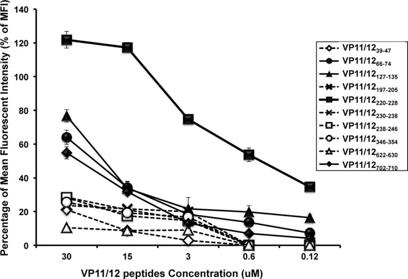
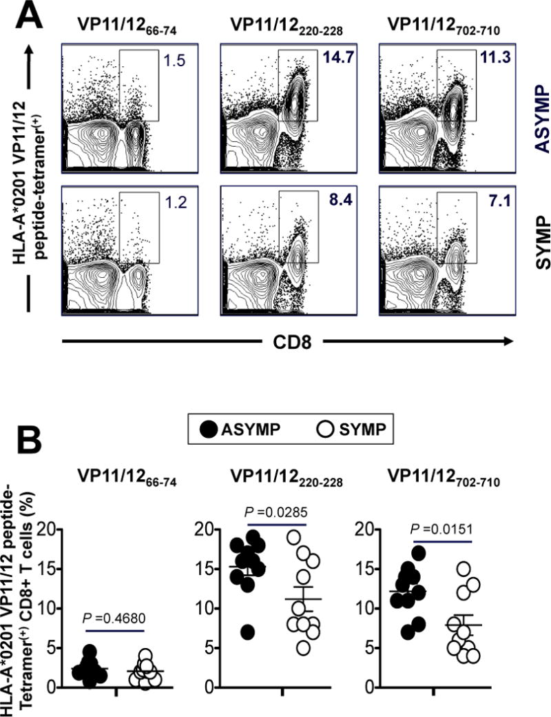
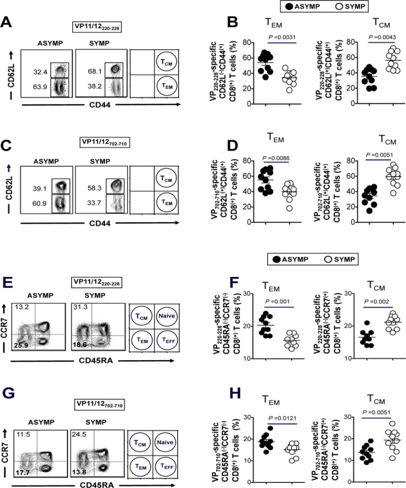
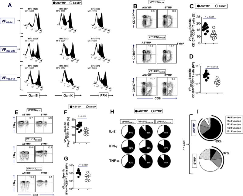
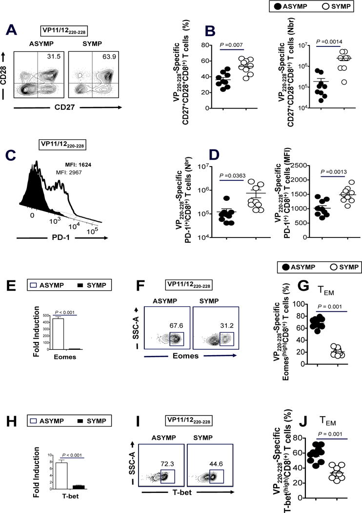
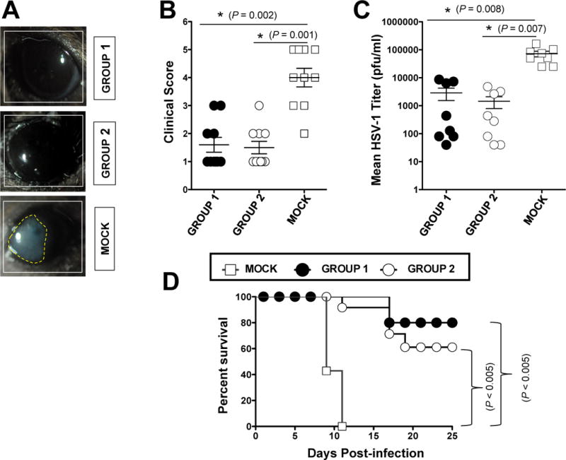
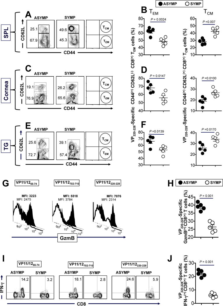
The HSV type 1 tegument virion phosphoprotein (VP) 11/12 (VP11/12) is a major Ag targeted by CD8(+) T cells from HSV-seropositive individuals. However, whether and which VP11/12 epitope-specific CD8(+) T cells play a role in the "natural" protection seen in seropositive healthy asymptomatic (ASYMP) individuals (who have never had clinical herpes disease) remain to be determined. In this study, we used multiple prediction computer-assisted algorithms to identify 10 potential HLA-A*02:01-restricted CD8(+) T cell epitopes from the 718-aa sequence of VP11/12. Three of 10 epitopes exhibited high-to-moderate binding affinity to HLA-A*02:01 molecules. In 10 sequentially studied HLA-A*02:01-positive and HSV-1-seropositive ASYMP individuals, the most frequent, robust, and polyfunctional effector CD8(+) T cell responses, as assessed by a combination of tetramer frequency, granzyme B, granzyme K, perforin, CD107(a/b) cytotoxic degranulation, IFN-γ, and multiplex cytokines assays, were predominantly directed against three epitopes: VP11/1266-74, VP11/12220-228, and VP11/12702-710. Interestingly, ASYMP individuals had a significantly higher proportion of CD45RA(low)CCR7(low)CD44(high)CD62L(low)CD27(low)CD28(low)CD8(+) effector memory CD8(+) T cells (TEMs) specific to the three epitopes, compared with symptomatic individuals (with a history of numerous episodes of recurrent ocular herpetic disease). Moreover, immunization of HLA-A*02:01 transgenic mice with the three ASYMP CD8(+) TEM cell epitopes induced robust and polyfunctional epitope-specific CD8(+) TEM cells that were associated with a strong protective immunity against ocular herpes infection and disease. Our findings outline phenotypic and functional features of protective HSV-specific CD8(+) T cells that should guide the development of an effective T cell-based herpes vaccine.


Conflict of interest statement

Conflict of interest: The authors have declared that no conflict of interest exists

Figures

Figure 1
Figure 1. Stabilization of HLA-A*0201 molecules by VP11/12 peptides on the surface of T2 cells
T2 cells (3 × 10⁵) were incubated with serial dilutions of the indicated VP11/12 peptide, as described in Materials & Methods. Cells were then stained with FITC conjugated anti-HLA-A2 mAb (BB7.2). The graph represents the percent of mean fluorescence intensity (MFI) reflecting an increase in the expression of HLA-A2 molecules on the surface of T2 cells triggered by various concentrations of VP11/12 peptides. The percent of MFI increase was calculated as follows: Percent of MFI increase = (MFI with the given peptide − MFI without peptide)/(MFI without peptide) × 100. Solid lines represent peptides that bind with high to moderate affinity to HLA-A02:01 molecules, as determined by stabilization of high levels of HLA-A*02:01 molecules on the surface of T2 cells when incubated with the indicated molarity of VP11/12 peptide. Broken lines represent low affinity peptide binders as determined by low levels of HLA-A*02:01 molecules stabilized on the surface of T2 cells. Error bars show standard deviation (SD) obtained from 3 independent experiments.
Figure 2
Figure 2. Frequencies of CD8+ T cells specific to VP11/12220–228 and VP11/12702–710 epitopes detected in HLA-A*02:01 positive HSV-seropositive ASYMP individuals compared to SYMP individuals
PBMCs (~10 × 106) derived from ten HLA-A*02:01 positive HSV-1 seropositive ASYMP individuals and from ten HLA-A*02:01 positive HSV-1 seropositive SYMP individuals were analyzed ex vivo by FACS for the frequency of CD8+ T cells specific to three VP11/12 epitopes using HLA-A*0201 VP11/12 peptide-tetramer complexes representing each of the three high to medium peptide binders (VP11/1266–74, VP11/12220–228 and VP11/12702–710 epitopes), as shown in Fig. 1. (A) Representative FACS data of the frequencies of CD8+ T cells, specific to VP11/1266–74, VP11/12220–228 and VP11/12702–710 epitopes, detected in PBMCs from one HLA-A*02:01 positive HSV-1 seropositive ASYMP individual (top panels) and one HLA-A*02:01 positive HSV-1 seropositive SYMP individual (lower panels). (B) Average frequencies of PBMC-derived CD8+ T cells, specific to VP11/1266–74, VP11/12220–228 and VP11/12702–710 epitopes, detected from ten HLA-A*02:01 positive HSV-1 seropositive ASYMP individuals compared to ten HLA-A*02:01 positive HSV-seropositive SYMP individuals. The results are representative of 2 independent experiments in each individual. The indicated P values, calculated using one-way ANOVA Test, show statistical significance between SYMP and ASYMP individuals.
Figure 3
Figure 3. Frequent VP11/12-epitope specific CD44highCD62LlowCD8+ TEM cells detected in ASYMP individuals compared to SYMP individuals
The phenotype of CD8+ T cells specific to VP11/12 peptide/Tetramer complexes representing each of the two immunodominant VP11/12220–228 and VP11/12702–710 epitopes were analyzed in term of TEM and TCM cells. (A) Representative FACS data of the frequencies of CD44highCD62LlowCD8+ TEM cells and CD44highCD62LhighCD8+ TCM cells specific to VP11/12220–228 peptide/Tetramer complexes from one HLA-A*02:01 positive HSV-1 seropositive ASYMP individual and from one HLA-A*02:01 positive HSV-1 seropositive SYMP individual. (B) Average frequencies of blood-derived CD8+ T cells specific to VP11/12220–228 peptide/Tetramer complexes with either TEM or TCM phenotype analyzed from 10 ASYMP and 10 SYMP individuals. (C) Representative data of the frequencies of CD44highCD62LlowCD8+ TEM and CD44highCD62LhighCD8+ TCM cells specific to VP11/12702–710 epitope from one ASYMP individual and one SYMP individual. (D) Average frequencies of blood-derived CD8+ T cells specific to VP11/12702–710 epitope with either TEM or TCM phenotype analyzed from 10 ASYMP and 10 SYMP individuals. Representative FACS data of the frequencies of CD45RAlowCCR7lowCD8+ TEM cells and CD45RAlowCCR7highCD8+ TCM cells, specific to (E) VP11/12220–228 epitope and (F) VP11/12702–710 epitope, analyzed in one ASYMP individual and one SYMP individual. Average frequencies of PBMC-derived CD8+ T cells with either TEM or TCM phenotype, specific to (G) VP11/12220–228 epitope and (H) VP11/12702–710 epitope, analyzed from 10 ASYMP and 10 SYMP individuals. The results are representative of 2 independent experiments in each individual. The indicated P values, calculated using one-way ANOVA Test, show statistical significance between SYMP and ASYMP individuals.
Figure 4
Figure 4. Asymptomatic individuals had a significantly higher proportion of polyfunctional HSV-1 VP11/12-epitope-specific CD8+ T cells
(A) VP11/12 epitope-primed CD8+ T cells from ASYMP individuals express high level of lytic granules compared to CD8+ T cells from SYMP individuals. FACS was used to determine the level of expression of GzmB, GzmK and PFN on tetramer gated CD8+ T cells specific to VP11/1266–74, VP11/12220–228 and VP11/12702–710 epitopes, as described in Materials & Methods. Samples were acquired on BD LSR II and data analysis was performed using FlowJo. The numbers on the top of each histogram represent mean fluorescent intensity (MFI) depicting the level of expression of each cytotoxic molecule. Numbers in bold represent mean fluorescent intensity (MFI) of ASYMP individual. (B) Representative FACS data of VP11/1266–74, VP11/12220–228 and VP11/12702–710 epitope-specific CD107a/bhighCD8+ T cells from one ASYMP vs. one SYMP individual. Average percentage (C) and average absolute number (D) of VP11/12220–228 epitope-specific CD107a/bhighCD8+ T cells from 10 ASYMP and 10 SYMP individuals. (E) Representative FACS data of VP11/1266–74, VP11/12220–228 and VP11/12702–710 epitope-specific IFN-γhighCD8+ T cells from one ASYMP vs. one SYMP individual. Average percentage (F) and average absolute number (G) of VP11/12220–228 epitope-specific IFN-γhighCD8+ T cells from 10 ASYMP and 10 SYMP individuals. (H) Summary pie charts showing the average amount of each cytokine produced by CD8+ T-cells from ASYMP patients (n = 10, black) and SYMP patients (n = 8, white), as detected by Luminex assay. The average frequencies of cytokine-producing CD8+ T cells from SYMP and ASYMP individuals are shown inside each pie chart. (I) Each pie chart represents the overall mean of CD8+ T cell functions from 10 HLA-A*02:01-positive ASYMP and 10 SYMP individuals in responses to stimulation with either SYMP or ASYMP VP11/12 peptides. Each sector of the pie chart represents the number of CD8+ T cell functions produced.
Figure 5
Figure 5. Asymptomatic individuals had a significantly higher proportion of differentiated and functional VP11/12-epitope-specific CD27highCD28highPD-1lowCD8+ T cells
Representative (A) and average frequencies (B) of VP220–228 epitope-specific CD8+ T cells from 10 ASYMP and vs. 10 SYMP individuals that are CD27highCD28high. Representative (C) and average frequencies (D) of VP220–228 epitope-specific CD8+ T cells from 10 ASYMP and vs. 10 SYMP individuals that are PD-1high. (E and H) The expression patterns of T-bet and Eomes transcription factors was analyzed, at RNA level using RT-PCR, from total CD8+ T cells derived from either SYMP (open square) or ASYMP individuals (closed square). Representative FACS data of the percentage of Eomes (F) and T-bet (I) positive HSV-1 VP11/12220–228 epitope-specific CD8+ T cells, derived from one SYMP individual (right) and one ASYMP individual (left). Average frequencies of the expression patterns of Eomes (G) and T-bet (J) transcription factors analyzed by FACS at the protein level from gated VP11/12220–228-specific CD8+ TEM cells derived from 10 SYMP (open circles) and 10 ASYMP individuals (closed circles). The results are representative of 3 independent experiments in each individual. The indicated P values, calculated using one-way ANOVA Test, show statistical significance between SYMP and ASYMP individuals.
Figure 6
Figure 6. Protective immunity against ocular herpes infection and disease induced by immunodominant VP11/12 CD8+ TEM epitopes in “humanized” HLA transgenic mice
Three groups of age-matched male HLA Tg mice (n = 10 each) were immunized subcutaneously, on days 0 and 21, with a mixture of three human CD8+ TEM cell epitope peptides (VP11/66–74, VP11/12220–228 and VP11/12702–710) delivered either with a novel CD4+ T cell epitope (VP11/12129–143) emulsified in CpG1826 adjuvant (Group 1) or with the promiscuous CD4+ T-cell PADRE epitope peptide emulsified in CpG1826 adjuvant (Group 2). CpG1826 adjuvant alone was used as control (Group 3, mock-vaccinated). Two weeks after the final immunization, all animals were challenged ocularly with 2 × 10⁵ pfu of HSV-1 (strain McKrae). The eye disease was detected and scored two weeks after immunization as described in Material & Methods (A and B). Virus titrations were determined from eye swabs on day 7 post-infection, as described in Material & Methods (C). Survival was determined in a window of 30 days post-challenge, as described in Material & Methods (D). The results are representative of 2 independent experiments. The indicated P values, calculated using one-way ANOVA Test, show statistical significance between protected (ASYMP) and non-protected (SYMP) mice.
Figure 7
Figure 7. The corneas of protected HSV-1 infected “ASYMP” HLA transgenic mice contain frequent VP11/12-specific polyfunctional CD8+ TEM cells
(A) Representative FACS data of the frequencies of CD44highCD62LlowCD8+ TEM and CD44highCD62LlowCD8+ TCM cells specific to VP11/12220–228 peptide/Tetramer complexes detected the corneas of one protected “ASYMP” vs. one non-protected “SYMP” HLA Tg mice. The spleen, cornea, and trigeminal ganglia were assayed for CD8+ T cell responses on day 9 post-infection. (B) Average frequencies of VP11/12220–228 epitope-specific TCM and TEM CD8+ cells detected in the corneas of five protected ASYMP and vs. five non-protected SYMP HLA Tg mice. ASYMP mice had a clinical score of 1.0 in Fig. 6B. SYMP mice had a score of 5 or 4 in Fig. 6B). Representative (C) and average frequencies (D) of VP11/1266–74 epitope-specific TCM and TEM CD8+ cells detected in the cornea five protected ASYMP and vs. five non-protected SYMP HLA Tg mice. (E) Representative data of level of expression of GzmB on cornea-derived CD8+ T cells specific to VP11/66–74, VP11/12220–228 and VP11/12702–710 detected from protected “ASYMP” mice (black histogram) and non-protected “SYMP” mice (white histogram). (F) Average frequencies of GzmB(+)CD8+ T cells specific to VP11/66–74, VP11/12220–228 and VP11/12702–710 detected from the corneas of protected “ASYMP” mice (black circles) and non-protected “SYMP” mice (white circles). (G) Representative data of the % IFN-γ(+)CD8+ T cells specific to VP11/66–74, VP11/12220–228 and VP11/12702–710 detected from the corneas of protected “ASYMP” mice (right) and non-protected “SYMP” mice (left). (H) Average frequencies of IFN-γ(+)CD8+ T cells specific to VP11/66–74, VP11/12220–228 and VP11/12702–710 detected from the corneas of protected “ASYMP” mice (black circles) and non-protected “SYMP” mice (white circles). 1×10⁶ cells for each assay and the results are representative of 2 independent experiments. The indicated P values, calculated using one-way ANOVA Test, show statistical significance between SYMP and ASYMP mice.

References

    1. Dervillez X, Qureshi H, Chentoufi AA, Khan AA, Kritzer E, Yu DC, Diaz OR, Gottimukkala C, Kalantari M, Villacres MC, Scarfone VM, McKinney DM, Sidney J, Sette A, Nesburn AB, Wechsler SL, BenMohamed L. Asymptomatic HLA-A*02:01-restricted epitopes from herpes simplex virus glycoprotein B preferentially recall polyfunctional CD8+ T cells from seropositive asymptomatic individuals and protect HLA transgenic mice against ocular herpes. J Immunol. 2013;191:5124–5138. - PMC - PubMed
    1. Chentoufi AA, Zhang X, Lamberth K, Dasgupta G, Bettahi I, Nguyen A, Wu M, Zhu X, Mohebbi A, Buus S, Wechsler SL, Nesburn AB, BenMohamed L. HLA-A*0201-restricted CD8+ cytotoxic T lymphocyte epitopes identified from herpes simplex virus glycoprotein D. J Immunol. 2008;180:426–437. - PubMed
    1. Zhang X, Dervillez X, Chentoufi AA, Badakhshan T, Bettahi I, Benmohamed L. Targeting the genital tract mucosa with a lipopeptide/recombinant adenovirus prime/boost vaccine induces potent and long-lasting CD8+ T cell immunity against herpes: importance of MyD88. J Immunol. 2012;189:4496–4509. - PMC - PubMed
    1. Samandary S, Kridane-Miledi H, Sandoval JS, Choudhury Z, Langa-Vives F, Spencer D, Chentoufi AA, Lemonnier FA, BenMohamed L. Associations of HLA-A, HLA-B and HLA-C alleles frequency with prevalence of herpes simplex virus infections and diseases across global populations: implication for the development of an universal CD8+ T-cell epitope-based vaccine. Hum Immunol. 2014;75:715–729. - PMC - PubMed
    1. Kuo T, Wang C, Badakhshan T, Chilukuri S, BenMohamed L. The Challenges and Opportunities for the Development of a T-Cell Epitope-Based Herpes Simplex Vaccine. Vaccine. 2014;34:715–729. - PMC - PubMed

Publication types

MeSH terms