. 2015 May;27(5):923-33.
doi: 10.1016/j.cellsig.2015.01.009. Epub 2015 Jan 22.

A small molecule PAI-1 functional inhibitor attenuates neointimal hyperplasia and vascular smooth muscle cell survival by promoting PAI-1 cleavage

Affiliations

A small molecule PAI-1 functional inhibitor attenuates neointimal hyperplasia and vascular smooth muscle cell survival by promoting PAI-1 cleavage

Tessa M Simone et al. Cell Signal. 2015 May.

Abstract

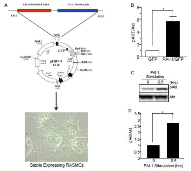
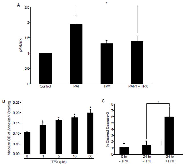
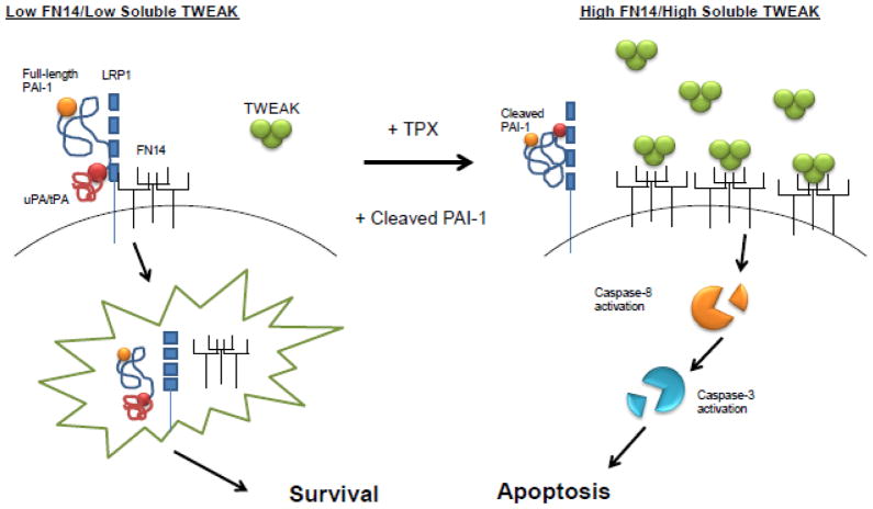
Plasminogen activator inhibitor-1 (PAI-1), the primary inhibitor of urokinase-and tissue-type plasminogen activators (uPA and tPA), is an injury-response gene implicated in the development of tissue fibrosis and cardiovascular disease. PAI-1 mRNA and protein levels were elevated in the balloon catheter-injured carotid and in the vascular smooth muscle cell (VSMC)-enriched neointima of ligated arteries. PAI-1/uPA complex formation and PAI-1 antiproteolytic activity can be inhibited, via proteolytic cleavage, by the small molecule antagonist tiplaxtinin which effectively increased the VSMC apoptotic index in vitro and attenuated carotid artery neointimal formation in vivo. In contrast to the active full-length serine protease inhibitor (SERPIN), elastase-cleaved PAI-1 (similar to tiplaxtinin) also promoted VSMC apoptosis in vitro and similarly reduced neointimal formation in vivo. The mechanism through which cleaved PAI-1 (CL-PAI-1) stimulates apoptosis appears to involve the TNF-α family member TWEAK (TNF-α weak inducer of apoptosis) and it's cognate receptor, fibroblast growth factor (FGF)-inducible 14 (FN14). CL-PAI-1 sensitizes cells to TWEAK-stimulated apoptosis while full-length PAI-1 did not, presumably due to its ability to down-regulate FN14 in a low density lipoprotein receptor-related protein 1 (LRP1)-dependent mechanism. It appears that prolonged exposure of VSMCs to CL-PAI-1 induces apoptosis by augmenting TWEAK/FN14 pro-apoptotic signaling. This work identifies a critical, anti-stenotic, role for a functionally-inactive (at least with regard to its protease inhibitory function) cleaved SERPIN. Therapies that promote the conversion of full-length to cleaved PAI-1 may have translational implications.

Keywords: Apoptosis; Carotid stenosis; PAI-1; SERPIN; Vascular injury.


Conflict of interest statement

6. Conflict of Interest

None declared.

Figures

Figure 1
Figure 1. PAI-1 expression is elevated in smooth muscle cells of the neointimal compartment of injured vessels
(A) RNA was extracted from balloon catheter-denuded and contralateral control carotid arteries one week following injury. Northern blots were hybridized using 32P-labeled cDNA probes to PAI-1 and A50 (loading control). (B) Paraffin sections (5 μm) of ligated (i–iii) and contralateral control (iv–vi) carotids were stained with H&E (i, iv) or processed for immunohistochemical detection of smooth muscle cell α-actin (ii, v) or PAI-1 (iii, vi) 14 days after common carotid artery ligation. Scale bar=100 μm.
Figure 2
Figure 2. Tiplaxtinin inhibits PAI-1/uPA complex formation and promotes PAI-1 cleavage
(A) Human uPA was incubated with FL-PAI-1 and the indicated concentration of tiplaxtinin. Solid arrow indicates full-length PAI-1; dashed arrow denotes cleaved PAI-1. (B) RASMCs were treated with tiplaxtinin or vehicle control in complete media for 24 h. PAI-1 and GAPDH (loading control) in cell lysates were identified by western blotting. Full-length and cleaved PAI-1 are highlighted by solid and dashed arrows, respectively. TPX=tiplaxtinin.
Figure 3
Figure 3. Vector-driven expression and exogenous addition of full-length PAI-1 stimulates Akt phosphorylation
(A) Schematic of a pEGFP-1-based vector in which a chimeric transcript consisting of 1.3 kb of PAI-1 coding sequences and GFP is expressed under the control of a 0.8 kb PAI-1 promoter. RASMCs were transfected with this PAI-1 coding sequence-GFP fusion construct under control of the PAI-1 promoter and stable expressing clones selected. (B) PAI-1 overexpression by 6- to 8-fold relative to GFP in RASMCs results in elevated pAkt levels. (C) PAI-1 addition induces a rapid and transient Akt activation in RASMCs. At confluence, RASMCs were serum-starved for 48 hours followed by stimulation with full-length PAI-1 (40 nM) for 30 minutes and cellular lysates analyzed by western blot. (D) pAkt levels were normalized to total Akt levels; histogram illustrates mean ± SEM; asterisk=p<0.05; n=3.
Figure 4
Figure 4. Tiplaxtinin inhibits PAI-1-induced Akt phosphorylation and promotes VSMC apoptosis
(A) RASMCs were serum-starved for 48 hours then treated with 40 nM PAI-1 with or without 10 nM tiplaxtinin. Cells were lysed, western blots probed for pAkt and normalized to Erk1. Data represent mean ± SEM, N=3. Asterisk=p ≤ 0.05; n=3. (B) RASMCs, cultured in 96-well plates, were treated with various concentrations of tiplaxtinin in complete media for 24 h prior to incubation of FITC-conjugated Annexin-V antibodies; apoptosis was measured using a fluorescence microplate reader. Data are presented as mean ± SEM; asterisks=p<0.05, n=3. (C) RASMCs were plated under reduced-serum conditions on tissue culture dishes pre-incubated with vitronectin and allowed to adhere and spread for 24 h. Cells were then either collected and fixed for immunocytochemical analysis of cleaved caspase-3 expression or treated with 10 μM tiplaxtinin or vehicle control for 24 h prior to fixation. Cleaved caspase-3-positive cells were scored as apoptotic and normalized to total cells (as assessed using DAPI staining). Data plotted are the percent mean ± SEM; asterisk=p<0.05, n=3. TPX=tiplaxtinin.
Figure 5
Figure 5. Elastase-cleaved PAI-1 promotes VSMC apoptosis while tiplaxtinin and CL-PAI-1 attenuate neointima formation
(A) Generation of CL-PAI-1 by incubation with human neutrophil elastase. Solid arrow indicates FL-PAI-1; dashed arrow denotes CL-PAI-1. (B) HuCASMCs treated with a final concentration of 10 nM FL-PAI-1 or CL-PAI-1 and a molar equivalent of elastase (as control) for 24 h were stained with ethidium homodimer and calcein AM to determine apoptotic index. Cells positive for ethidium homodimer were scored as apoptotic and normalized against calcein AM (total cells). Data are presented as mean ± SEM; asterisks=p<0.05, n=3. (C) HuCASMCs were treated with CL-PAI-1 (40 nM) and/or staruosporine (5 μM) as indicated for 24 h. Caspase-3 and actin (loading control) were assessed in whole cell lysates by western blot. (D) H&E stained cross-sections of contralateral control and ligated carotid arteries isolated from mice treated once daily with vehicle control (Con), 3 mg/kg tiplaxtinin (via oral gavage), 2 mg/kg FL-PAI-1 or CL-PAI-1 (both by intraperitoneal injection) for 14 days following ligation. Arrowheads indicate the internal elastic lamina. (E) Quantitation of the intima-to-media ration for the indicated ligated carotid artery groups. Data plotted represent the mean ± SEM; asterisks=p<0.05, n=4. (F) Time line of carotid ligation experiments; arrows indicate days of treatment with the indicated reagents.
Figure 6
Figure 6. Cleaved PAI-1 promotes VSMC apoptosis in a plasmin-dependent manner
(A) HuCASMCs were treated with tiplaxtinin or CL-PAI-1 for 6 h (left) or with the indicated siRNAs for 48 h (right). Plasminogen, plasmin, PAI-1 and GAPDH (loading control) were evaluated in cell lysates by western blotting. (B) HuCASMCs were incubated with 100 μM tranexamic acid for 30 min prior to the addition of 40 nM CL-PAI-1. Cleaved PARP and actin (loading control) were assessed in cell lysates. (C) RASMCs were pretreated with 1.25 μM plasminogen for 30 min prior to addition of tiplaxtinin (10 μM, final concentration) for 24 h. Cells were collected, stained with Annexin-V and analysed by FACS. TPX=tiplaxtinin; CL-PAI-1=cleaved PAI-1
Figure 7
Figure 7. Upregulation of caspase-8 activation, FN14 and soluble TWEAK in response to cleaved PAI-1 and tiplaxtinin
(A) HuCASMCs were incubated with tiplaxtinin or CL-PAI-1 in the concentrations indicated for 24 h. Whole cell extracts were separated by gel electrophoresis and blots probed with antibodies to caspase-8 and actin (loading control). Bands: 56 kDa=full-length caspase-8; 43, 41 kDa= cleaved caspase-8; 18 kDa= cleaved cleaved caspase-8; ns=non-specific band. (B) HuCASMCs were treated with 40 nM FL-PAI-1, 40 nM CL-PAI-1 or 10 μM tiplaxtinin for 24 hours, extracted and western blots developed using antibodies to TWEAK (depicted in table in B as mean ± SEM normalized to Erk2 expression, n=3) and FN14 using Erk2 as a loading control; NS=not significant. (C) Histogram is a plot of the mean ± SEM of the conditioned media levels of soluble TWEAK (relative to Ponceau S-stained total protein), n=4. TPX=tiplaxtinin; FL-PAI-1=full length PAI-1; CL-PAI-1=cleaved PAI-1
Figure 8
Figure 8. Full-length PAI-1 down-regulates FN14 in an LRP1-dependent manner and protects against TWEAK-stimulated apoptosis
(A) HuCASMCs were treated with 40 nM FL- PAI-1 or CL-PAI-1 for 24 h. Surface FN14 expression was assessed by FACS. Graphed in (A) is a plot of the mean ± SEM; asterisks=p<0.05, n=4. Below the graph is a representative FACS histogram. n/s indicates no statistical significance. (B) PAI-1 induction of pAkt is LRP1-dependent. RASMCs were serum-starved for 48 hours before a 5 minute pretreatment with 5 μg/ml RAP prior to PAI-1 stimulation for 30 minutes. Cells were lysed and pAkt/Erk levels assessed by western blot analysis; pAkt levels were normalized to Erk1 and represented as a mean ± SEM, n=3. (C) LRP1−/− cells were treated with vehicle alone or 40 nM PAI-1 for 30 minutes; lysates were analyzed by western blot analysis. pAkt levels were normalized to total Erk1 levels. Data is represented as the mean ± SEM, n=3. Asterisks = p <0.05. n/s indicates no statistical significance. (D) RASMCs were incubated with 40 nM R76E-PAI-1 for 24 h, a recombinant PAI-1 mutant deficient in LRP1-binding. FACS was used to quantify surface FN14 expression. Graphed in (D) is a plot of the mean ± SEM; asterisks=p<0.05, n=4. n/s indicates no statistical significance. Below the graph is a representative FACS histogram. (E) HuCASMC were treated with 40 nM FL- PAI-1, CL-PAI-1 or 40 nM R76E-PAI-1 and 250 ng/ml soluble TWEAK, or TWEAK alone, for 6 h. Cells were collected, lysed and an aminomethylcoumarin (AMC)-tagged caspase-3 target (Z-DEVD) added. Fluorescence emission was monitored using a microplate reader every hour for 24 h. Data plotted represents the mean fluorescent intensity over a 24-h period ± SEM; asterisks=p<0.05 using a repeated-measures ANOVA. TPX=tiplaxtinin; FL-PAI-1=full length PAI-1; CL-PAI-1=cleaved PAI-1; R76E-PAI-1=LRP1-binding deficient mutant recombinant PAI-1 protein
Figure 9
Figure 9. Hypothetical model
Current findings suggest that PAI-1 regulates VSMC survival and potentiates neointimal formation. Full-length PAI-1 binds to, and inhibits, uPA attenuating the conversion of plasminogen to plasmin while simultaneously down-regulating surface FN14 receptor expression through LRP1-mediated endocytosis, promoting a pro-survival, pro-stenotic phenotype. When the conformational pools of PAI-1 shift to increased levels of cleaved PAI-1 (either by tiplaxtinin or exogenous addition), conversion of plasminogen to plasmin is amplified. Membrane-bound and soluble TWEAK expression is increased stimulating, thereby, FN14-TWEAK signaling leading to VSMC apoptosis and reduced neointimal hyperplasia.

References

    1. Bochaton-Piallat ML, Gabbiani F, Redard M, Desmouliere A, Gabbiani G. Apoptosis participates in cellularity regulation during rat aortic intimal thickening. Am J Pathol. 1995;146:1059–1064. - PMC - PubMed
    1. Schwartz SM. Perspectives series: cell adhesion in vascular biology. Smooth muscle migration in atherosclerosis and restenosis. J Clin Invest. 1997;99:2814–2816. - PMC - PubMed
    1. Declerck PJ, De Mol M, Vaughan DE, Collen D. Identification of a conformationally distinct form of plasminogen activator inhibitor-1, acting as a noninhibitory substrate for tissue-type plasminogen activator. J Biol Chem. 1992;267:11693–11696. - PubMed
    1. Urano T, Strandberg L, Johansson LB, Ny T. A substrate-like form of plasminogen-activator-inhibitor type 1. Conversions between different forms by sodium dodecyl sulphate. Eur J Biochem. 1992;209:985–992. - PubMed
    1. Dellas C, Loskutoff DJ. Historical analysis of PAI-1 from its discovery to its potential role in cell motility and disease. Thromb Haemost. 2005;93:631–640. - PubMed

MeSH terms