. 2015 Jan 24;10(1):e0116784.
doi: 10.1371/journal.pone.0116784. eCollection 2015.

Detrusor induction of miR-132/212 following bladder outlet obstruction: association with MeCP2 repression and cell viability

Affiliations

Detrusor induction of miR-132/212 following bladder outlet obstruction: association with MeCP2 repression and cell viability

Mardjaneh Karbalaei Sadegh et al. PLoS One. .

Abstract

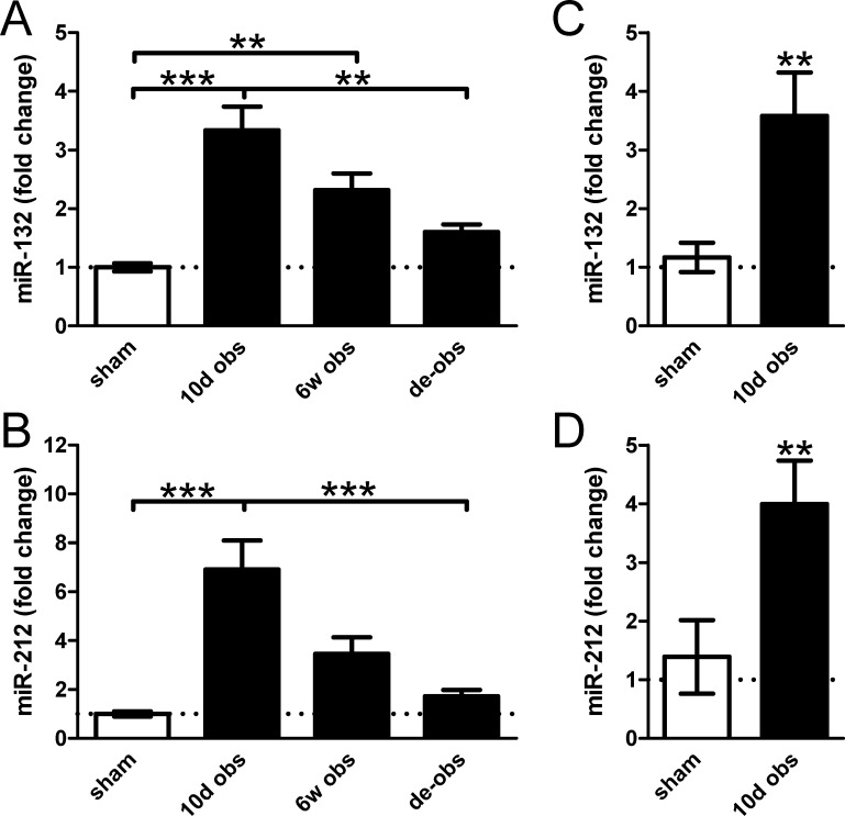
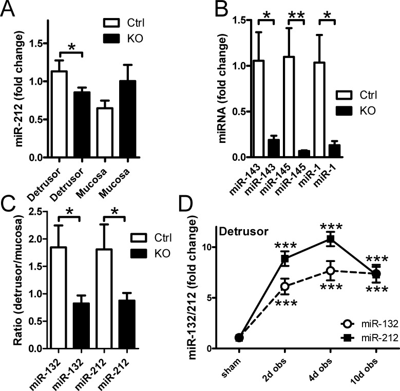
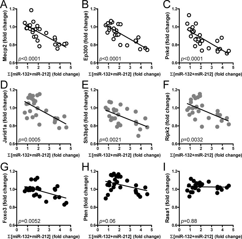
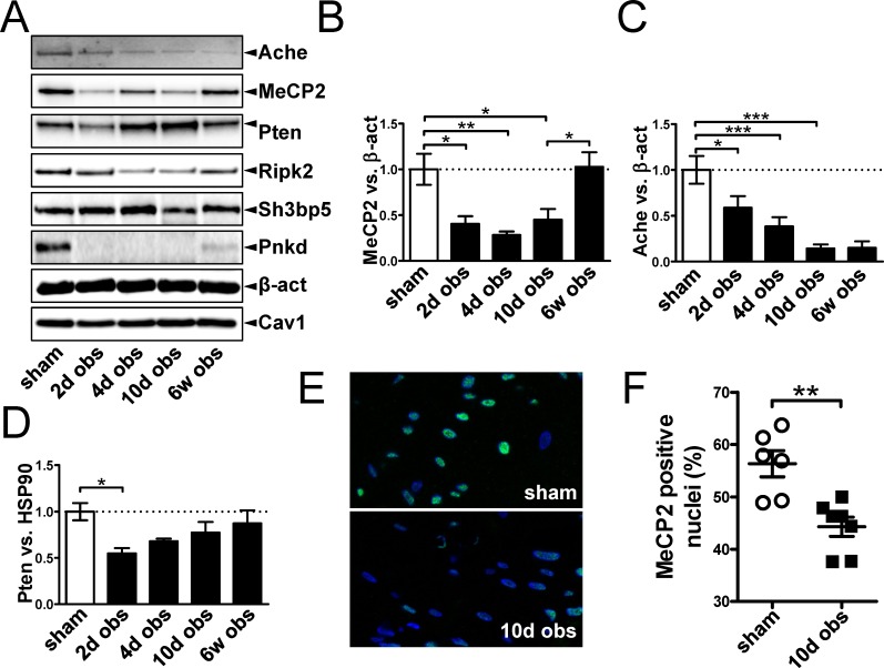
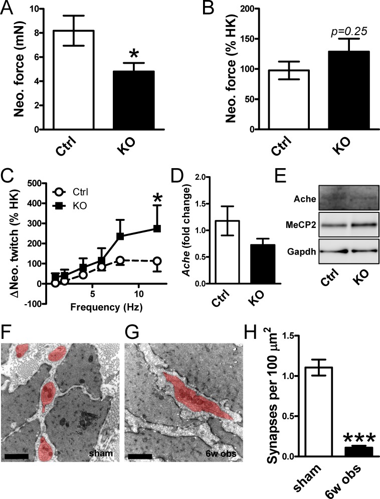
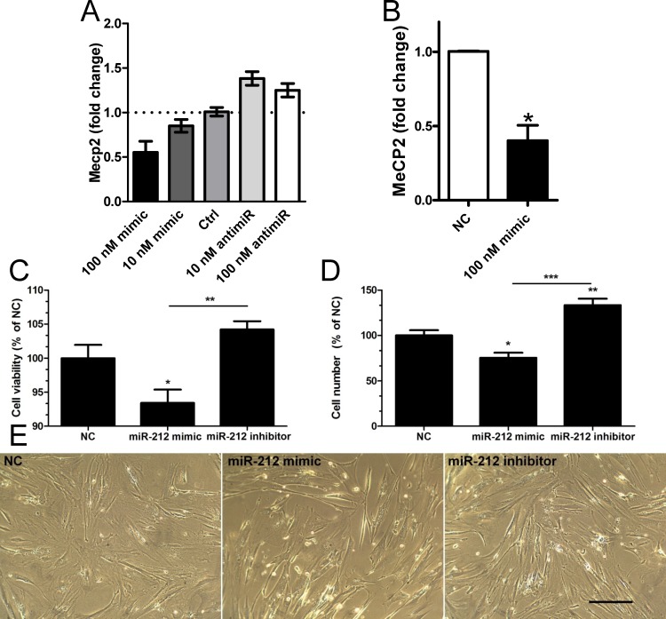
The microRNAs (miRNAs) miR-132 and miR-212 have been found to regulate synaptic plasticity and cholinergic signaling and recent work has demonstrated roles outside of the CNS, including in smooth muscle. Here, we examined if miR-132 and miR-212 are induced in the urinary bladder following outlet obstruction and whether this correlates with effects on gene expression and cell growth. Three to seven-fold induction of miR-132/212 was found at 10 days of obstruction and this was selective for the detrusor layer. We cross-referenced putative binding sites in the miR-132/212 promoter with transcription factors that were predicted to be active in the obstruction model. This suggested involvement of Creb and Ahr in miR-132/212 induction. Creb phosphorylation (S-133) was not increased, but the number of Ahr positive nuclei increased. Moreover, we found that serum stimulation and protein kinase C activation induced miR-132/212 in human detrusor cells. To identify miR-132/212 targets, we correlated the mRNA levels of validated targets with the miRNA levels. Significant correlations between miR-132/212 and MeCP2, Ep300, Pnkd and Jarid1a were observed, and the protein levels of MeCP2, Pnkd and Ache were reduced after obstruction. Reduction of Ache however closely matched a 90% reduction of synapse density arguing that its repression was unrelated to miR-132/212 induction. Importantly, transfection of antimirs and mimics in cultured detrusor cells increased and decreased, respectively, the number of cells and led to changes in MeCP2 expression. In all, these findings show that obstruction of the urethra increases miR-132 and miR-212 in the detrusor and suggests that this influences gene expression and limits cell growth.


Conflict of interest statement

Competing Interests: The authors have declared that no competing interests exist.

Figures

Figure 1
Figure 1. Partial infravesical outlet obstruction increases expression of miR-132/212 in the urinary bladder.
Rats were sham-operated (sham) or subjected to obstruction for 10 days (10d obs) and 6 weeks (6w obs). One group of rats were obstructed for 6 weeks and then re-operated to remove the obstruction. The obstruction-induced hypertrophy was then allowed to regress for another 10 days (de-obs). Bladders were harvested, RNA was isolated and microarrays were run (GEO accession GSE47080). Results in panels A and B are from these microarrays. Panels C and D show confirmation using qRT-PCR at 10 days of obstruction versus sham. *, **, and *** denote p<0.05, p<0.01 and p<0.001 in this and the following figures. N = 6–8 throughout.
Figure 2
Figure 2. Basal expression of miR-132/212 in detrusor smooth muscle is low yet induction following outlet obstruction occurs only in the detrusor layer.
Control mice and mice with smooth muscle-specific deletion of Dicer (KO) were used to examine the contribution of smooth muscle cells to miR-132 and miR-212 expression in the bladder (panels A through C). Panel A shows miR-212 expression in control and KO mice following separation of the detrusor and mucosa using micro-dissection. Panel B shows expression of miR-143, miR-145 and miR-1 in control and KO detrusor. Panel C shows the detrusor to mucosa expression ratio of miR-132 and miR-212 in control and KO bladders. Time-courses of miR-132/212 expression in detrusor following partial bladder outlet obstruction in the rat are shown in panel D. N = 4–10 throughout.
Figure 3
Figure 3. Bioinformatics analysis points to the involvement of Ahr in miR-132/212 induction following outlet obstruction.
Transcription factor binding site (TFBS) analysis identified significant enrichment of 93 transcription factor binding motifs in promoters of differentially expressed genes at 10 days of obstruction (blue circle in A). These were cross-referenced against putative transcription factor binding sites in the miR-132/212 promoter (red circle in A), yielding a list of putative mediators of miR-132/212 induction in outlet obstruction (B). Hits with low expression levels and those with raw p-values exceeding 0.01 are shown in thin lettering. Panel C shows phosphorylation of CREB (S133) in bladders from sham-operated rats and following various time of obstruction. Panel D shows immunofluorescence labeling (green) of the dioxin receptor Ahr in bladders from sham-operated and obstructed rats. Dotted lines indicate the outer surface of the urinary bladder. The scale bar to the left applies to all images and represents 100 μm. Panels E through G show miR-132 expression in human bladder smooth muscle cells stimulated with vehicle (control) and various pharmacological substances in vitro. TCDD: 2,3,7,8-tetrachlorodibenzo-p-dioxin; FCS: fetal calf serum; PMA: Phorbol 12-myristate 13-acetate. Further details are given in Materials and Methods.
Figure 4
Figure 4. MiR-132/212 induction in the bladder correlates inversely with previously validated miR-132/212 targets.
The normalized sum of miR-132 and miR-212 in bladders from sham-operated, obstructed (10 days and 6 weeks) and de-obstructed rats was correlated with the mRNA levels for Mecp2 (A), Ep300 (B), Pnkd (C), Jarid1a (D), Sh3bp5 (E), Ripk2 (F), Foxo3 (G), Pten (H) and Rasa1 (I). Expression data were obtained from microarray experiments (GEO accession GSE47080). Each symbol represents one rat and the 10d obstructed rats are represented by the rightmost symbols. The p-values obtained for the individual correlations are given at the bottom left in each diagram.
Figure 5
Figure 5. Reduced levels of the miR-132/212 targets Ache, MeCp2 and Pnkd in the bladder following outlet obstruction.
Validated targets of miR-132/212 were examined at the protein level using western blotting at various times after surgical obstruction of the urethra (A). Summarized data (n = 6) for MeCP2, Ache and Pten is shown in panels B through D. Panel E shows double staining for MeCP2 (green) and DNA (blue) in control and obstructed (10 days) bladders. Panel F shows the percentage of MeCP2 positive nuclei in bladders from sham-operated and obstructed (10 days) rats (n = 6 and 7, respectively).
Figure 6
Figure 6. Smooth muscle-specific deletion of Dicer does not increase neostigmine-induced contraction or expression of Ache in the bladder.
Panels A and B show contraction induced by the addition of the acetylcholine-esterase (Ache) inhibitor neostigmine in control and in Dicer knockout (KO) bladder strips. Force is given in mN (A) and relative to depolarization-induced contraction (B), respectively. Panel C shows the potentiation by neostigmine of twitches in response to electrical field stimulation (ΔNeo. twitch) in control and Dicer KO detrusor strips. Panels D and E show the mRNA and protein levels of Ache in control and Dicer KO bladders. Panels F and G show representative electron micrographs from sham-operated and obstructed (6 weeks) bladders. Neural varicosities are highlighted in red. Scale bars represent 1 μm. Panel H shows quantitative evaluation of synapse density in control and obstructed bladders. N = 6.
Figure 7
Figure 7. Transfection of miR-132/212 mimics and inhibitors affect MeCP2 expression and cell number in vitro.
An initial dosing experiment performed in duplicate using human detrusor smooth muscle cells from one individual shows a reciprocal effect of miR-132 mimic and inhibitor on MeCP2 expression (A). The effect of 100 nM miR-132 mimic was confirmed using cells from three individuals (B). Panel C shows effect of miR-212 mimic and inhibitor (100 nM of each) on cell viability using the MTT assay. Panel D shows the effect of miR-212 mimic and inhibitor on cell number and panel E shows phase contrast light microscopic images of cells transfected with negative control (NC), miR-212 mimic and miR-212 inhibitor. The scale bar in E indicates 20 μm and n = 5 and 8 in C and D, respectively.

References

    1. Filipowicz W, Bhattacharyya SN, Sonenberg N (2008) Mechanisms of post-transcriptional regulation by microRNAs: are the answers in sight? Nat Rev Genet 9: 102–114. 10.1038/nrg2290 - DOI - PubMed
    1. Lee Y, Ahn C, Han J, Choi H, Kim J, et al. (2003) The nuclear RNase III Drosha initiates microRNA processing. Nature 425: 415–419. 10.1038/nature01957 - DOI - PubMed
    1. Sadegh MK, Ekman M, Rippe C, Uvelius B, Sward K, et al. (2012) Deletion of Dicer in smooth muscle affects voiding pattern and reduces detrusor contractility and neuroeffector transmission. PLoS One 7: e35882 10.1371/journal.pone.0035882 - DOI - PMC - PubMed
    1. Ekman M, Bhattachariya A, Dahan D, Uvelius B, Albinsson S, et al. (2013) Mir-29 repression in bladder outlet obstruction contributes to matrix remodeling and altered stiffness. PLoS One 8: e82308 10.1371/journal.pone.0082308 - DOI - PMC - PubMed
    1. Wanet A, Tacheny A, Arnould T, Renard P (2012) miR-212/132 expression and functions: within and beyond the neuronal compartment. Nucleic Acids Res 40: 4742–4753. 10.1093/nar/gks151 - DOI - PMC - PubMed

Publication types

MeSH terms