Review
. 2015 May;25(5):276-85.
doi: 10.1016/j.tcb.2014.12.008. Epub 2015 Jan 21.

Ciliary ectosomes: transmissions from the cell's antenna

Affiliations
Review

Ciliary ectosomes: transmissions from the cell's antenna

Christopher R Wood et al. Trends Cell Biol. 2015 May.

Abstract

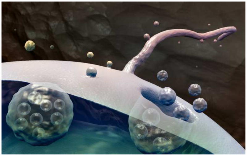
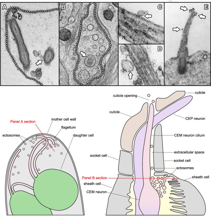
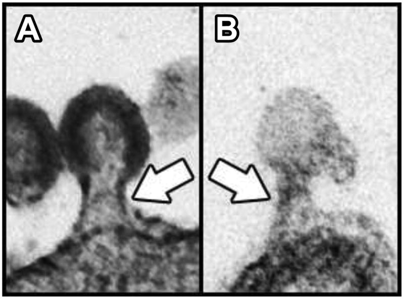
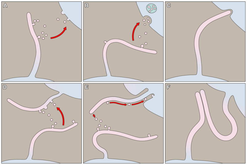
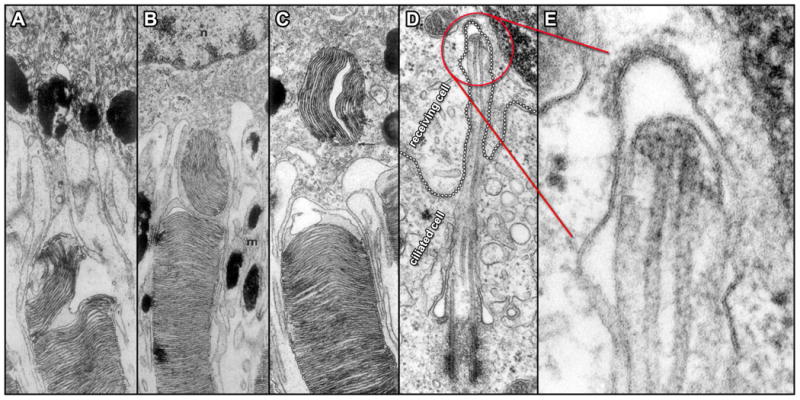
The cilium is the site of function for a variety of membrane receptors, enzymes and signal transduction modules crucial for a spectrum of cellular processes. Through targeted transport and selective gating mechanisms, the cell localizes specific proteins to the cilium that equip it for the role of sensory antenna. This capacity of the cilium to serve as a specialized compartment where specific proteins can be readily concentrated for sensory reception also makes it an ideal organelle to employ for the regulated emission of specific biological material and information. In this review we present and discuss an emerging body of evidence centered on ciliary ectosomes - bioactive vesicles released from the surface of the cilium.

Keywords: cilium; extracellular vesicles; intercellular signaling.


Figures

Figure 1
Figure 1
The release of extracellular vesicles. Exosomes are released when multivesicular bodies fuse with the cell membrane. Ectosomes are released by outward budding directly from the cell membrane or cilium membrane. (illustration by Christopher R. Wood)
Figure 2
Figure 2
Observations of extracellular vesicles and cilia. A. TEM of an ultrathin section through a mature Chlamydomonas sporangium shows ectosomes in the extracellular space surrounding flagella. The location of the mother cell wall is indicated by a dotted line and an arrow indicates ectosomes caught in the process of budding directly from the flagellar membrane (). B. A tomographic cross-section through a C. elegans cephalic sensillum shows numerous ectosomes (arrowheads) populating the extracellular space around a CEM neuronal cilium (arrow). The boundary of the sensillar lumen is indicated by a dotted line (). C, D. TEMs of ultrathin sections through mouse biliary primary cilia show extracellular vesicles (arrows) closely apposed to the ciliary membranes. () E. TEM of an ultrathin section through the primary cilium of a mouse neuroepithelial cell shows prominin-1 specific gold particles decorating what appear to be regions of the ciliary membrane in the process of forming ectosomes (arrows) (, courtesy of Michaela Wilsch-Bräuninger). A cartoon at the lower left illustrates a region of a Chlamydomonas sporangium like the one sectioned in panel A. Two flagellated daughter cells are shown releasing ciliary ectosomes into the extracellular space within the lumen of the mother cell wall. The red line indicates the orientation of the section depicted in panel A. A cartoon at the lower right illustrates a region of a cephalic male sensillum (CEM) like the one sectioned in panel B. Glial sheath and socket cells form a lumen surrounding the CEM neuron cilium. Ectosomes are found in the luminal space and may travel along the cilium to the cuticle opening where they are released into the worm's external environment. The red line indicates the orientation of the section depicted in panel B.
Figure 3
Figure 3
Comparison between a viral bud and a ciliary ectosomal bud. A. TEM of an ultrathin section through an HIV-1 particle arrested in the process of budding from the plasma membrane of a human cell in culture (). B. TEM of an ultrathin section through a ciliary ectosome caught in the process of budding from the membrane of a Chlamydomonas flagellum (). White arrows indicate the appearance of similar electron-dense structures within the two bud necks.
Figure 4
Figure 4
Modes of ciliary membrane interaction. A. Ciliary ectosomes may fuse with the plasma membrane of a recipient cell. B. Ciliary ectosomes may be endocytosed by a recipient cell. C. A cilium may interact with a recipient cell by direct membrane contact. D. Extracellular vesicles may fuse with the ciliary membrane of a recipient cell. E. Extracellular vesicles may adhere to the ciliary membrane of a recipient cell and be moved along the length of the cilium by sub-membrane motor activity. F. A cilium may interact with another cilium by direct membrane contact.
Figure 5
Figure 5
Direct interactions between cilia and neighboring cell surfaces. A – C. TEMs of ultrathin sections through three different rod outer segment tips illustrate the sequential events in the interaction with adjacent RPE cells in Rhesus monkey. A grouping of outer segment disks initially undergoes separation within the outer segment (A); followed by engulfment via RPE cell cytoplasmic extensions (B) and movement deeper into the cytoplasm of the RPE cell (C) (). D. TEM of an ultrathin section through a fibroblast from the testes of rat shows the cell membrane of a receiving cell (emphasized by a dotted outline) forming what appears to be a coated pit in response to an intruding primary cilium extending from an adjacent ciliated cell. Panel E displays a higher magnification view of the ciliary tip region seen in D and indicated in red ().

References

    1. Pazour GJ, et al. Chlamydomonas IFT88 and its mouse homologue, polycystic kidney disease gene tg737, are required for assembly of cilia and flagella. J Cell Biol. 2000;151:709–718. - PMC - PubMed
    1. Pazour GJ, Rosenbaum J. Intraflagellar transport and cilia-dependent diseases. Trends Cell Biol. 2002;12:551–555. - PubMed
    1. Pazour GJ, Witman GB. The vertebrate primary cilium is a sensory organelle. Curr Opin Cell Biol. 2003;15:105–10. - PubMed
    1. Rosenbaum JL, Witman GB. Intraflagellar Transport. Nature Rev Cell Mol Biol. 2002;3:813–825. - PubMed
    1. Badano JL, Mitsuma N, Beales PL, Katsanis N. The ciliopathies: an emerging class of human genetic disorders. Annu Rev Genomics Hum Genet. 2006;7:125–48. - PubMed

LinkOut - more resources