. 2015 Jan 26:5:8026.
doi: 10.1038/srep08026.

Structure of mycobacterial maltokinase, the missing link in the essential GlgE-pathway

Affiliations

Structure of mycobacterial maltokinase, the missing link in the essential GlgE-pathway

Joana Fraga et al. Sci Rep. .

Abstract

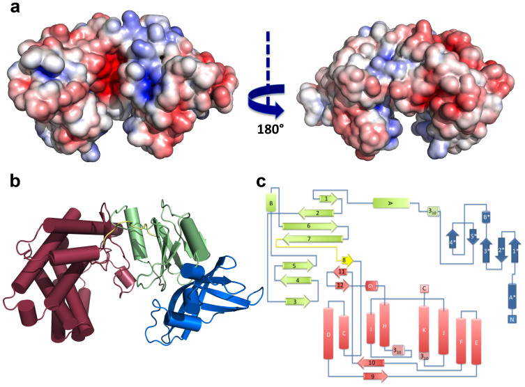
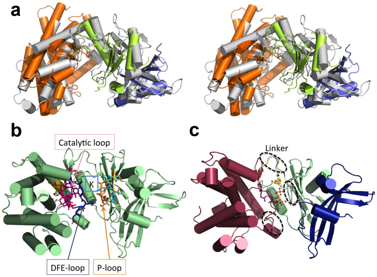
A novel four-step pathway identified recently in mycobacteria channels trehalose to glycogen synthesis and is also likely involved in the biosynthesis of two other crucial polymers: intracellular methylglucose lipopolysaccharides and exposed capsular glucan. The structures of three of the intervening enzymes - GlgB, GlgE, and TreS - were recently reported, providing the first templates for rational drug design. Here we describe the structural characterization of the fourth enzyme of the pathway, mycobacterial maltokinase (Mak), uncovering a eukaryotic-like kinase (ELK) fold, similar to methylthioribose kinases and aminoglycoside phosphotransferases. The 1.15 Å structure of Mak in complex with a non-hydrolysable ATP analog reveals subtle structural rearrangements upon nucleotide binding in the cleft between the N- and the C-terminal lobes. Remarkably, this new family of ELKs has a novel N-terminal domain topologically resembling the cystatin family of protease inhibitors. By interfacing with and restraining the mobility of the phosphate-binding region of the N-terminal lobe, Mak's unusual N-terminal domain might regulate its phosphotransfer activity and represents the most likely anchoring point for TreS, the upstream enzyme in the pathway. By completing the gallery of atomic-detail models of an essential pathway, this structure opens new avenues for the rational design of alternative anti-tubercular compounds.


Figures

Figure 1
Figure 1. Maltokinase distribution in the bacterial domain.
Maximum likelihood phylogenetic tree, built using 350 bacterial species and a concatenated alignment of 31 highly conserved protein-coding genes for phylogenetic inference (adapted by permission from Macmillan Publishers Ltd: Nature, copyright 2009). Bacterial phyla and type of Mak occurrence are identified by different colours.
Figure 2
Figure 2. Biochemical characterization of MakMvan.
Influence of buffer pH (A) and temperature (B) on MakMvan enzymatic activity. (C) Specificity of MakMvan for different phosphate donor substrates. (D) Effect of divalent cations on enzyme activity. Plotted data are the mean of three independent experiments.
Figure 3
Figure 3. Overall structure of MakMvan.
(A) Solid-surface representation of MakMvan, with mapped electrostatic surface potential contoured from +5 (blue) to −5 (red) kbTe−1 [kb, Boltzmann's constant; T, temperature (K); e, charge of an electron]. The left and right views are related by a 180° rotation around the vertical axis. (B) Cartoon representation of MakMvan three-dimensional structure, coloured with the distinct domains in different colors (cap domain: blue; intermediate domain: green; C-terminal domain: pink). (C) Topology diagram of MakMvan, color-coded as in panel B. Panels A and B were prepared with PyMOL (http://www.pymol.org).
Figure 4
Figure 4. MakMvan is structurally similar to eukaryotic protein kinase-like proteins (ELKs).
(A) Stereoview of MakMvan structure (grey cartoon) superposed with MTRK (PDB entry 2PUL32) N-terminal (green) and C-terminal (orange) domains, and with the cystatin domain (blue) of multicystatin (PDB entry 2W9P63). (B) Mapping of ELK structural motifs onto MakMvan three-dimensional structure (P-loop: orange; AMLKV motif: cyan; catalytic loop with HGD motif: magenta; DFE motif: blue; DVA motif: light pink; VAXVH motif: yellow). (C) Cartoon representation of MakMvan:AppCp complex structure depicting the subtle structural changes required to accommodate the nucleotide (dashed ellipsoids). The bound nucleotide (for simplicity, only one of the two modelled conformations is shown) is represented as ball-and-stick (carbon: yellow; nitrogen: blue; oxygen red; phosphorous: orange), magnesium ions are shown as spheres (magenta) and the magnesium-coordinating residues (Gln310 and Asp322) are represented as sticks (atom color as in nucleotide except for carbon atoms that are coloured pink). Figures prepared with PyMOL (http://www.pymol.org).
Figure 5
Figure 5. Close view of the AppCp-binding site of MakMvan.
(A) Stereo view of the polar interactions centred on the phosphoryl moieties of AppCp. For all panels, residues are coloured according to conservation as shown in Supplementary Fig. S1, where red corresponds to positions strictly conserved in the actinobacteria maltokinase sequences selected for the multiple alignment. Selected residues are shown as sticks with oxygen atoms in red, nitrogen in blue, sulfur in yellow, phosphorus in orange and carbon in green (nucleotide) or according to sequence conservation (protein). The putative catalytic base (Asp305) is represented as ball-and-stick. Hydrogen bonds are represented as dashed black lines. The magnesium ions are represented as magenta spheres and dashed magenta lines represent bonds to the magnesium coordinating atoms. Red spheres represent ordered water molecules. (B) Detailed view of the interactions with the adenine base of the nucleotide bound to the active site cleft of MakMvan. (C) Close-up of the vicinity of residue Asn137, which bridges the P-loop and the terminal phosphoryl moiety of AppCp to the N-terminal cap domain through the conserved Arg152. Figure prepared with PyMOL (http://www.pymol.org).
Figure 6
Figure 6. Identification of surface cavities in MakMvan.
(A) Surface representation of free MakMvan coloured according to residue conservation (consistent with the multiple sequence alignment shown in Supplementary Fig. S1). Cavities identified with the software fpocket and mentioned in the main text are represented as dotted spheres (pocket 1: blue; pocket 2: cyan; pocket 3: white; pocket 4: light green; pocket 5: light pink). (B) Close view of the residues lining pocket 1 coloured as in panel A. Residue Asp305 is represented as ball-and-stick and highlighted by a dashed blue line; the γ-phosphate of AppCp (ball-and-stick representation) is highlighted by a blue line. (C) Detailed view of residues delimiting pocket 2, coloured as in panel A; Asp305 is represented as ball-and-stick and circled by a dashed line. (D) Close view of the cavity located at the interface between the intermediate (green) and the N-terminal cap domain (blue); Asn137 is highlighted by a rectangle and Arg152 by a dashed ellipse. Hydrogen bonds are represented as dashed lines. For panels B–D, important residues are shown as sticks with oxygen atoms in red, nitrogen in blue, sulfur in yellow, phosphorus in orange and carbon in green (nucleotide) or according to sequence conservation (protein). Panels C–D refer to the structure of MakMvan in complex with AppCp. Figure prepared with PyMOL (http://www.pymol.org).

References

    1. Sambou T. et al. Capsular glucan and intracellular glycogen of Mycobacterium tuberculosis: biosynthesis and impact on the persistence in mice. Mol Microbiol 70, 762–774, 10.1111/j.1365-2958.2008.06445.x (2008). - DOI - PMC - PubMed
    1. Chandra G., Chater K. F. & Bornemann S. Unexpected and widespread connections between bacterial glycogen and trehalose metabolism. Microbiology 157, 1565–1572, 10.1099/mic.0.044263-0 (2011). - DOI - PubMed
    1. Kalscheuer R. et al. Self-poisoning of Mycobacterium tuberculosis by targeting GlgE in an alpha-glucan pathway. Nat Chem Biol 6, 376–384, 10.1038/nchembio.340 (2010). - DOI - PMC - PubMed
    1. Elbein A. D., Pastuszak I., Tackett A. J., Wilson T. & Pan Y. T. Last step in the conversion of trehalose to glycogen: a mycobacterial enzyme that transfers maltose from maltose 1-phosphate to glycogen. J Biol Chem 285, 9803–9812, 10.1074/jbc.M109.033944 (2010). - DOI - PMC - PubMed
    1. Kalscheuer R. & Jacobs W. R. Jr The significance of GlgE as a new target for tuberculosis. Drug News Perspect 23, 619–624, 10.1358/dnp.2010.23.10.1534855 (2010). - DOI - PubMed

Publication types

MeSH terms

Substances

LinkOut - more resources