. 2015 Aug;35(4):354-89.
doi: 10.1111/neup.12189. Epub 2015 Jan 26.

Arizona Study of Aging and Neurodegenerative Disorders and Brain and Body Donation Program

Affiliations

Arizona Study of Aging and Neurodegenerative Disorders and Brain and Body Donation Program

Thomas G Beach et al. Neuropathology. 2015 Aug.

Abstract

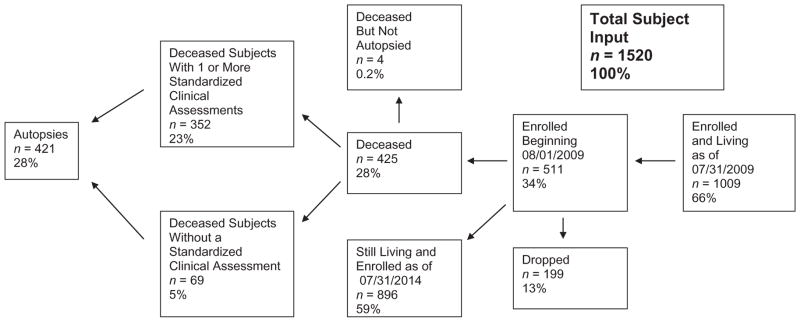
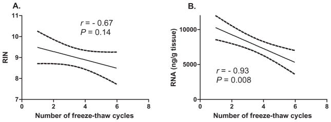
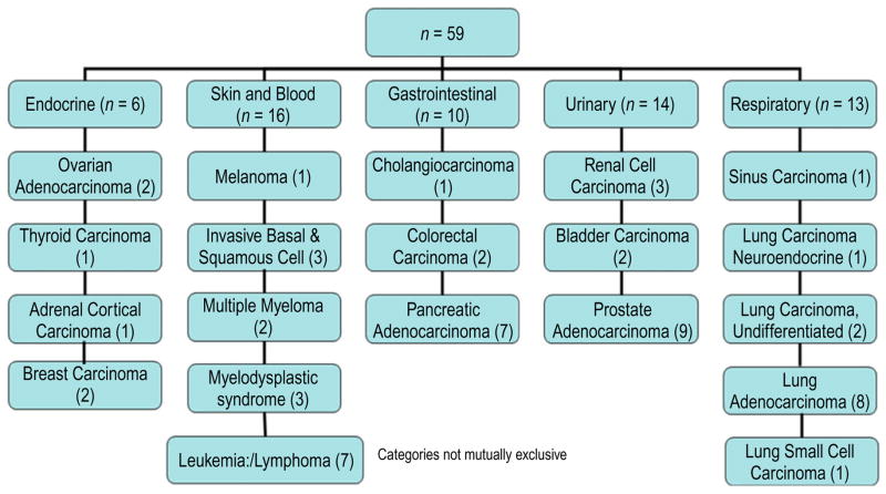
The Brain and Body Donation Program (BBDP) at Banner Sun Health Research Institute (http://www.brainandbodydonationprogram.org) started in 1987 with brain-only donations and currently has banked more than 1600 brains. More than 430 whole-body donations have been received since this service was commenced in 2005. The collective academic output of the BBDP is now described as the Arizona Study of Aging and Neurodegenerative Disorders (AZSAND). Most BBDP subjects are enrolled as cognitively normal volunteers residing in the retirement communities of metropolitan Phoenix, Arizona. Specific recruitment efforts are also directed at subjects with Alzheimer's disease, Parkinson's disease and cancer. The median age at death is 82. Subjects receive standardized general medical, neurological, neuropsychological and movement disorders assessments during life and more than 90% receive full pathological examinations by medically licensed pathologists after death. The Program has been funded through a combination of internal, federal and state of Arizona grants as well as user fees and pharmaceutical industry collaborations. Subsets of the Program are utilized by the US National Institute on Aging Arizona Alzheimer's Disease Core Center and the US National Institute of Neurological Disorders and Stroke National Brain and Tissue Resource for Parkinson's Disease and Related Disorders. Substantial funding has also been received from the Michael J. Fox Foundation for Parkinson's Research. The Program has made rapid autopsy a priority, with a 3.0-hour median post-mortem interval for the entire collection. The median RNA Integrity Number (RIN) for frozen brain and body tissue is 8.9 and 7.4, respectively. More than 2500 tissue requests have been served and currently about 200 are served annually. These requests have been made by more than 400 investigators located in 32 US states and 15 countries. Tissue from the BBDP has contributed to more than 350 publications and more than 200 grant-funded projects.

Keywords: Alzheimer's disease; Parkinson's disease; RNA; aging; autopsy; biobank; biospecimen; brain bank; cancer; freeze-thaw; pathology; post-mortem interval.


Figures

Fig 1
Fig 1
Autopsied subjects of the Brain and Body Donation Program (BBDP) 08/01/2009–07/30/2014. Percentages are calculated with respect to total subject input. Of autopsied subjects, 84% had one or more standardized clinical assessments.
Fig 2
Fig 2
Correlation between the number of thawing cycles and (A) RNA Integrity Number (RIN) and (B) RNA yield, measured as the concentration (ng/μL) per tissue weight (g). Dashed lines show outlines of 95% confidence intervals.
Fig. 3
Fig. 3
Photomicrographs illustrating the histological methods used for investigating neurodegenerative diseases. Frames A–D are 40 μm large format sections stained with Campbell-Switzer silver stain to show all types of senile plaques. Frames E–H are 40 μm large format sections stained with Gallyas silver stain, showing both neurofibrillary changes (neurofibrillary tangles, dystrophic neurites and neuropil threads) and neuritic plaques. Frames I–M show, in 40 μm large format sections, changes of progressive supranuclear palsy, including neurofibrillary tangles in the substantia nigra (I), pontine nuclei (K) and dentate nucleus (L, M), and tufted astrocytes (J). Frame J was obtained from a section stained with the Gallyas method while I and K–M were from sections stained with an immunoperoxidase method for phosphorylated tau protein (AT8 antibody). Frames N and O show, in 40 μm large format sections, astrocytic plaques in a subject with corticobasal degeneration. Frames P–R show changes of Pick’s disease, including a chromatolytic “Pick cell” (P, H, E paraffin section), Pick bodies in the dentate granule cell layer (Q, 40 μm large format section stained with the Campbell-Switzer method) and oligodendroglial coiled bodies (R, 40 μm large format section stained with the Gallyas method). Frame S shows argyrophilic grains in a 40 μm large format section stained with the Gallyas method. Section T is a low-magnification image of the amgydala and adjacent temporal lobe, in a subject with dementia with Lewy bodies, from a 40 μm large format section stained with an immunoperoxidase method for phosphorylated α-synuclein. Frames U–Y are from subjects with Parkinson’s disease (PD) or dementia with Lewy bodies (DLB), stained with an immunoperoxidase method for phosphorylated α-synuclein. Frames U, V and X are from paraffin sections (U – olfactory bulb; V – locus ceruleus; X – submandibular gland). Frame W is from an 80 μm large format tangential section of the lower esophagus. Frame Y is from a retinal whole-mount from a PD subject. Frame Z is from a 40 μm large format section, from a subject with frontotemporal lobar dementia, stained with an immunoperoxidase method for phosphorylated TDP-43.
Fig 4
Fig 4
Photographs and photomicrographs illustrating vascular lesions. Frame A shows cross-sections of the circle of Willis from cognitively normal subjects with relatively normal arteries (above) as compared to late-onset Alzheimer’s disease subjects with relatively severe atherosclerotic stenosis (below). Frames B–F are from 40 μm large format sections. Frame B shows periventricular white matter rarefaction in an HE-stained section of the frontal lobe. Frame C shows a microinfarct in the cerebral cortex while frame D shows a microscopic focus of cerebellar cortical sclerosis, both from HE-stained sections. Frame E shows amyloidotic capillaries in the primary visual cortex, stained with the Campbell-Switzer silver stain. Frame F shows a larger amyloidotic blood vessel with dyshoric diffuse perivascular amyloid, stained with the thioflavin S method.
Fig 5
Fig 5
Frozen cancer tissue types available. All have detailed clinical and pathological data. Many have both primary site and metastatic tissue samples.

References

    1. Beach TG. Alzheimer’s disease and the “Valley of Death”: not enough guidance from human brain tissue? J Alzheimers Dis. 2013;33 (Suppl 1):S219–S233. - PubMed
    1. Peschken CA, Esdaile JM. Rheumatic diseases in North America’s indigenous peoples. Semin Arthritis Rheum. 1999;28:368–391. - PubMed
    1. Johanneson B, Steinsson K, Lindqvist AK, et al. A comparison of genome-scans performed in multicase families with systemic lupus erythematosus from different population groups. J Autoimmun. 1999;13:137–141. - PubMed
    1. Lio D, Pes GM, Carru C, et al. Association between the HLA-DR alleles and longevity: a study in Sardinian population. Exp Gerontol. 2003;38:313–317. - PubMed
    1. Mathews CA, Reus VI, Bejarano J, et al. Genetic studies of neuropsychiatric disorders in Costa Rica: a model for the use of isolated populations. Psychiatr Genet. 2004;14:13–23. - PubMed

Publication types