

doi: 10.1042/BST20140324.
The myosin mesa and a possible unifying hypothesis for the molecular basis of human hypertrophic cardiomyopathy
Affiliations
- PMID: 25619247
- PMCID: PMC4349527
- DOI: 10.1042/BST20140324
Item in Clipboard
The myosin mesa and a possible unifying hypothesis for the molecular basis of human hypertrophic cardiomyopathy
Biochem Soc Trans.
2015 Feb.
Abstract
No matter how many times one explores the structure of the myosin molecule, there is always something new to discover. Here, I describe the myosin mesa, a structural feature of the motor domain that has the characteristics of a binding domain for another protein, possibly myosin-binding protein C (MyBP-C). Interestingly, many well-known hypertrophic cardiomyopathy (HCM) mutations lie along this surface and may affect the putative interactions proposed here. A potential unifying hypothesis for the molecular basis of human hypertrophic cardiomyopathy is discussed here. It involves increased power output of the cardiac muscle as a result of HCM mutations causing the release of inhibition by myosin binding protein C.
Figures

(A) Chick embryo fibroblasts extracted with the detergent triton-X 100. The ‘cytoskeleton’ is revealed. (B) A dividing Dictyostelium myosin-II null cell in which GFP-tagged myosin was inserted to rescue cytokinesis. The amount of myosin moving to the cleavage furrow is quantified by the distribution of fluorescence intensity. (C) A human induced pluripotent stem (iPS)-cell-derived cardiomyocyte stained with phalloidin (actin, red) and a β-cardiac myosin-specific antibody (yellow). Image by Arjun Adhikari, Alexandre Ribeiro and Kristina Bezold.

MyBP-C is known to interact with both the myosin thick filament and the actin filament. Shown are Tm, Tn and S1, the globular head domain of myosin, which contains the myosin light chains (not shown) and is the motor domain of the molecule.

(A) A side view showing the catalytic domain oriented to illustrate the flat mesa (dashed oval), with the actin-binding region on the left and the light-chain-binding lever arm on the right. The N-terminal 25 K (light grey), 50 K (white) and C-terminal 20 K (medium grey) domains are shown, as well as the two light chains (dark grey) that are part of the lever arm. The two residues that mark the beginning and end of loop 1 (cyan) are near the edge of the mesa. (B) The mesa viewed from the top (dashed oval; view in A rotated toward you 90°). The two subdomains of the 50 K domain are marked. Loop 2 (not shown in the crystal structure) is a prominent feature of the mesa, the beginning and end of which is marked by the cyan residues. The structure shown here, and in all figures below, is a chimera of the human β-cardiac catalytic domain (MYH7; through to the converter) crystal structure (pdb id: 4DB1) with the converter and lever arm modelled in from the chicken skeletal myosin structure (MYH2) (pdb id: 2MYS). (C) The mesa viewed at an oblique angle and in surface electrostatic mode (large dashed oval). Basic patches are more raised than neighboring acidic patches. A particularly large positive cluster very rich in arginine residues (small dashed oval) is near loop 1. The beginning residues of Loops 1 and 2 (cyan) are shown. Images here and in figures below were rendered in PyMOL.

(A) A view of the cartoon form of S1, looking at the face of the molecule nearly opposite to the mesa surface. The red residues mark major changes in the molecule. They are found throughout the S1; changes in the light-chain-binding region are not shown. The cyan residues mark loops 1 and 2. (B) The myosin mesa viewed from the top. Note the absence of major changes on this surface.

(A) Human β-cardiac myosin (MYH7). (B) Major changes (orange residues) when compared with human skeletal muscle myosin (MYH2). (C) Major changes (red residues) when compared with human smooth muscle myosin (B1PS43). (D) Major changes (red residues) when compared with human non-muscle myosin II (MYH10).
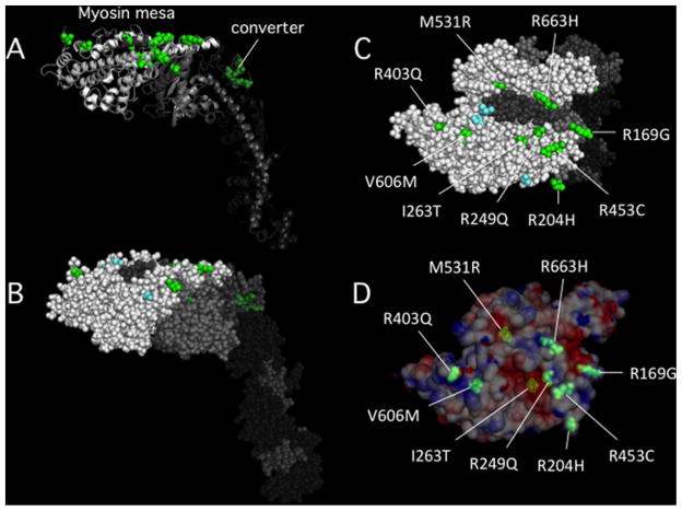
(A) Cartoon image view showing two groups of HCM mutations. Four mutations are in the converter domain (dark green) and 11 are on or very near the myosin mesa (bright green). Fourteen of the mutations were chosen because they have been well-documented to be the cause of HCM in families carrying these mutations. The fifteenth is M531R, which, although only documented in one family, is a left ventricular non-compaction mutant myosin that appears to be hypercontractile in our studies. (B) The same view as in (A) except in all sphere representation. (C) The top view of the mesa showing that nine of the 11 mutations are on the mesa surface (the other two are just below the surface). (D) The same view as in (C) except the surface charge distribution is shown. M531R and I263T are slightly below the surface in acidic pockets, while the remainder of the mutations are right at the surface and most are arginine residues, 5 of which form a particularly large domain of positive charge on the lower right (see also Figure 3C, small dashed oval).

(A) Cartoon image of DCM mutations (orange) showing the same view as in Figure 6A. Most are not on the myosin mesa. The DCM mutations were chosen on the basis of high likelihood of being causative of DCM [45]. They are I201T, A223T, R237W, G245E, I248F, R369Q, D469Y, I524V, E525K, S532P, I533V, R567H, N597K and F764L. (B) The view from the other side of the S1 compared to the view in A. (C) The top view of the mesa showing that 4 of the 14 mutations are on the mesa surface, and all in a clustered linear sequence. (D) The same view as in C except the surface charge distribution is shown.
References
-
- Spudich JA. How molecular motors work. Nature. 1994;372:515–518. - PubMed
-
- Spudich JA. The myosin swinging cross-bridge model. Nat Rev Mol Cell Biol. 2001;2:387–392. - PubMed
-
- Brown S, Levinson W, Spudich JA. Cytoskeletal elements of chick embryo fibroblasts revealed by detergent extraction. J Supramol Struct. 1976;5:119–130. - PubMed
Publication types
MeSH terms
Substances
Grants and funding
LinkOut - more resources
Full Text Sources