. 2015 Apr;19(4):734-43.
doi: 10.1111/jcmm.12387. Epub 2015 Jan 26.

Persistent DNA damage-induced premature senescence alters the functional features of human bone marrow mesenchymal stem cells

Affiliations

Persistent DNA damage-induced premature senescence alters the functional features of human bone marrow mesenchymal stem cells

Valentina Minieri et al. J Cell Mol Med. 2015 Apr.

Abstract

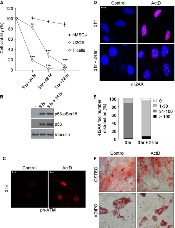
Human mesenchymal stem cells (hMSCs) are adult multipotent stem cells located in various tissues, including the bone marrow. In contrast to terminally differentiated somatic cells, adult stem cells must persist and function throughout life to ensure tissue homeostasis and repair. For this reason, they must be equipped with DNA damage responses able to maintain genomic integrity while ensuring their lifelong persistence. Evaluation of hMSC response to genotoxic insults is of great interest considering both their therapeutic potential and their physiological functions. This study aimed to investigate the response of human bone marrow MSCs to the genotoxic agent Actinomycin D (ActD), a well-known anti-tumour drug. We report that hMSCs react by undergoing premature senescence driven by a persistent DNA damage response activation, as hallmarked by inhibition of DNA synthesis, p21 and p16 protein expression, marked Senescent Associated β-galactosidase activity and enlarged γH2AX foci co-localizing with 53BP1 protein. Senescent hMSCs overexpress several senescence-associated secretory phenotype (SASP) genes and promote motility of lung tumour and osteosarcoma cell lines in vitro. Our findings disclose a multifaceted consequence of ActD treatment on hMSCs that on the one hand helps to preserve this stem cell pool and prevents damaged cells from undergoing neoplastic transformation, and on the other hand alters their functional effects on the surrounding tissue microenvironment in a way that might worsen their tumour-promoting behaviour.

Keywords: DNA damage; actinomycin D; mesenchymal stem cell; senescence-associated secretory phenotype; stress-induced premature senescence.


Figures

Fig 1
Fig 1
hMSCs are resistant to ActD-induced DNA damage. (A) Survival of ActD-treated cells was analysed by PI staining and flow cytometry. Cells were treated for 3 hrs with ActD (400 mM) and allowed to recovery in drug-free medium for 24, 48 and 72 hrs. Values were normalized on control cells and represented as mean ± SD of three independent experiments. Student's T-test was performed and the significance values indicated by asterisks. (B) Immunoblotting of total and phosphoSer15-p53 in hMSCs after 3 hrs of ActD treatment and after 24 hrs of recovery. Vinculin was used as loading control; sign (-) indicates control cells. (C) Immunofluorescence of ATM autophosphorylation in Ser1981 (ph-ATM) in hMSCs after 3 hrs of ActD treatment. (D) Immunofluorescence of γH2AX (red) foci induction in hMSCs after 3 hrs of ActD treatment and of residual foci after 24 hrs of recovery. DAPI counterstains the nuclei (blue). (E) γH2AX foci number quantification in ActD-treated hMSCs; one representative experiment out of two is shown. (F) Control and ActD-treated hMSCs were induced to differentiate after 24 hrs of recovery. Top panel: Alizarin Red S staining for calcium deposition after 21 days’ culture. 10× magnification images were taken with a contrast phase microscope. Bottom panel: Oil Red staining for lipid accumulation after 15 days’ culture. 20× magnification images were taken with a contrast phase microscope. In all panels the white bar represents 10 μm.
Fig 2
Fig 2
hMSCs activate SIPS following ActD treatment. (A) Percentage of EdU positive cells on the total Hoechst 33342 positive cells at 1 and 9 days of recovery after 3 hrs of ActD treatment. Mean ± SEM of three independent experiments is shown. Student's t-test was performed and the significance value indicated by asterisks. (B) Immunoblotting of p53, p21 and p16 proteins at 0, 1, 2, 3, 6 and 9 days of recovery. β-Actin was used as loading control. Sign (-) indicates control cells. (C) Representative images of β-galactosidase staining after 21 days of recovery (phase contrast microscope 10× magnification images). (D) Immunofluorescence analysis of persistent γH2AX foci (red) after 21 days of recovery. DAPI counterstains the nuclei (blue). (E) Immunofluorescence co-localization analysis of γH2AX (green) and 53BP1 (red) after 21 days of recovery. In all panels the white bar represents 10 μm.
Fig 3
Fig 3
Senescent hMSCs augment the expression of inflammatory cytokine genes and promote tumour cell migration. (A) qPCR relative gene expression levels of nine SASP factors in ActD-treated hMSCs after 9 and 15 days of recovery. Relative variation of transcript levels in ActD-treated hMSCs was expressed as Log2 (fold change), calculated by using Actβ as reference gene and time-matched control cells as calibrator. One representative experiment out of two. (B) Influence of conditioned medium obtained from senescent and control hMSCs on the migration ability of U2OS and CALU-1 cell lines. Shown are representative images (bottom) and graph (top) of migrating cell number/field (mean ± SD) derived from three independent migration assays. Student's t-test was performed and the significance values indicated by asterisks. (C) Influence of conditioned medium obtained from senescent and control hMSCs on the proliferation of U2OS and CALU-1 cell line. CellTiter-Blue fluorescence values were normalized on control cells and represented as mean ± SD of three independent experiments.

References

    1. Pittenger MF, Mackay AM, Beck SC, et al. Multilineage potential of adult human mesenchymal stem cells. Science. 1999;284:143–7. - PubMed
    1. Frenette PS, Pinho S, Lucas D, et al. Mesenchymal stem cell: keystone of the hematopoietic stem cell niche and a stepping-stone for regenerative medicine. Annu Rev Immunol. 2013;31:285–316. - PubMed
    1. Nery AA, Nascimento IC, Glaser T, et al. Human mesenchymal stem cells: from immunophenotyping by flow cytometry to clinical applications. Cytometry A. 2013;83:48–61. - PubMed
    1. Satija NK, Singh VK, Verma YK, et al. Mesenchymal stem cell-based therapy: a new paradigm in regenerative medicine. J Cell Mol Med. 2009;13:4385–402. - PMC - PubMed
    1. Sperka T, Wang J, Rudolph KL. DNA damage checkpoints in stem cells, ageing and cancer. Nat Rev Mol Cell Biol. 2012;13:579–90. - PubMed

Publication types

MeSH terms