. 2015 Feb 3;23(2):332-41.
doi: 10.1016/j.str.2014.10.025. Epub 2015 Jan 22.

The molecular basis for histone H4- and H2A-specific amino-terminal acetylation by NatD

Affiliations

The molecular basis for histone H4- and H2A-specific amino-terminal acetylation by NatD

Robert S Magin et al. Structure. .

Abstract

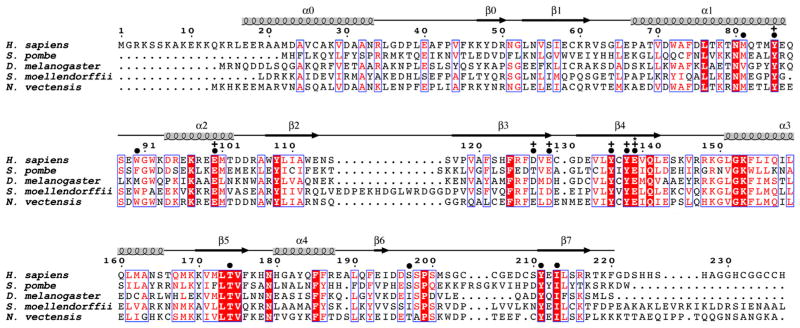
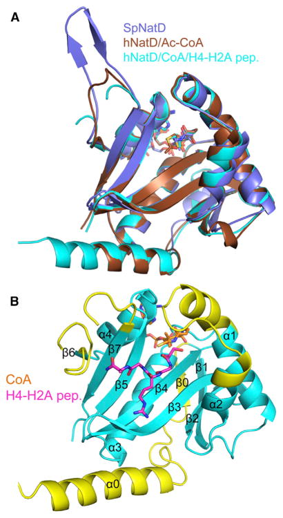
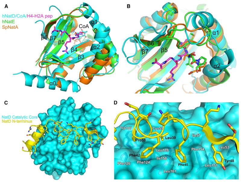
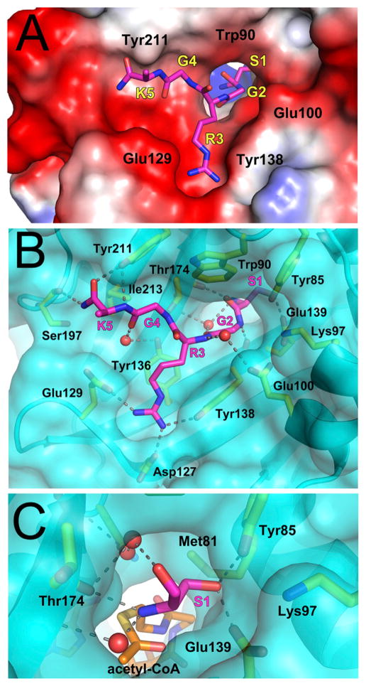
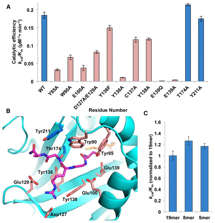
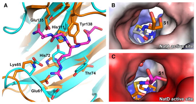
N-terminal acetylation is among the most common protein modifications in eukaryotes and is mediated by evolutionarily conserved N-terminal acetyltransferases (NATs). NatD is among the most selective NATs; its only known substrates are histones H4 and H2A, containing the N-terminal sequence SGRGK in humans. Here we characterize the molecular basis for substrate-specific acetylation by NatD by reporting its crystal structure bound to cognate substrates and performing related biochemical studies. A novel N-terminal segment wraps around the catalytic core domain to make stabilizing interactions, and the α1-α2 and β6-β7 loops adopt novel conformations to properly orient the histone N termini in the binding site. Ser1 and Arg3 of the histone make extensive contacts to highly conserved NatD residues in the substrate binding pocket, and flanking glycine residues also appear to contribute to substrate-specific binding by NatD, together defining a Ser-Gly-Arg-Gly recognition sequence. These studies have implications for understanding substrate-specific acetylation by NAT enzymes.


Figures

Figure 1
Figure 1. Sequence Alignment of NatD Orthologs
The sequence alignment contains the following NatD orthologs: human (Homo sapiens), fission yeast (Schizosaccharomyces pombe), fruit fly (Drosophila melanogaster), spikemoss (Selaginella moellendorffii), and sea anemone (Nematostella vectensis). The blue boxes represent conserved patches of sequence alignment. Residues in red are highly conserved, and residues in white with a red background are strictly conserved. Above the sequence alignment are indicated amino acid numbering for H. sapiens and secondary structure elements. Amino acid residues are indicated that make contacts to the substrate peptide (●), show mutational sensitivity (+), or are proposed to play catalytic roles (*).
Figure 2
Figure 2. Overall Structure of NatD Complexes
(A) Superposition of the SpNatD/acetyl-CoA (violet), hNatD/acetyl-CoA (brown), and hNatD/CoA/H4-H2A peptide (cyan) complexes. CoA is shown as sticks, and the H4-H2A peptide is omitted for clarity. (B) Overall structure of hNatD/CoA/H4-H2A peptide with structurally unique elements of NatD relative to other NATs highlighted in yellow. CoA is shown in orange and H4 is shown in magenta.
Figure 3
Figure 3. Unique Structural Features of NatD
(A) Superposition of hNatD (cyan), SpNatA (orange), and hNatE (green) complexes. The H4-H2A peptide is shown in magenta. Only the CoA from the hNatD/CoA/H4-H2A peptide structure is shown for clarity. (B) Close-up view of the substrate binding groove of NatD in comparison with NatA and NatE. The color coding is as in Figure 3A. NatA substrate (SASE) and NatE substrate (MLGP) are shown as orange and green sticks, respectively. (C) View of the interaction of the NatD N-terminal segment (yellow cartoon representation) with the catalytic core domain (cyan surface representation). (D) Detailed interactions between the N-terminal segment and catalytic core domain of NatD. Residues from the N terminus that mediate interactions are labeled in black, and residues from the core domain are colored in dark blue and labeled in white. Met-162 is omitted for clarity.
Figure 4
Figure 4. Peptide Binding Site of NatD
(A) Electrostatic surface of the NatD peptide binding site with the peptide shown in magenta stick figure. Residues from NatD are labeled in black and residues from the peptide are labeled in yellow with their corresponding one-letter codes and numerical positions in the peptide. The side chain of Lys5p was disordered and not modeled into the crystal structure. (B) Detailed interactions between NatD and the H4-H2A peptide. NatD is shown as a transparent cyan surface, and residues that interact with the peptide are yellow. Hydrogen bonds are shown as dashed lines and waters are shown as red spheres. (C) Close-up view of the NatD active site highlighting interactions made by Ser1p. Acetyl-CoA was modeled into the figure by aligning the binary and ternary NatD structures.
Figure 5
Figure 5. Mutational Analysis of NatD
(A) Catalytic efficiency of selected NatD mutants. Mutations that have negligible effect are in blue, while those that decrease the catalytic efficiency of the enzyme are in pink. (B) Residues targeted for mutagenesis are mapped onto the NatD structure. The color scheme is the same as Figure 5A. (C) The catalytic efficiency of wild-type NatD toward N-terminal histone H4 peptides of varying length. Data are represented as mean ± SEM. Each mutant and peptide of varying length was assayed in triplicate.
Figure 6
Figure 6. Comparison between Substrate Recognition of NatD and NatA
(A) Overlay of the peptide binding site of NatA (orange) and NatD (cyan with magenta histone substrate). (B) Electrostatic potential surface of the NatA active site with a bound covalently linked bisubstrate inhibitor. The N-terminal serine of the substrate peptide is shown in orange, and the acetyl-CoA moiety of the bisubstrate inhibitor is in white. (C) Electrostatic potential surface of the NatD active site. The N-terminal serine of the substrate peptide is shown in magenta, and acetyl-CoA is in orange. Waters are shown as red spheres. Acetyl-CoA was modeled into the figure by aligning the binary and ternary NatD structures.

References

    1. Adams PD, Afonine PV, Bunkoczi G, Chen VB, Echols N, Headd JJ, Hung LW, Jain S, Kapral GJ, Grosse Kunstleve RW, et al. The Phenix software for automated determination of macromolecular structures. Methods. 2011;55:94–106. - PMC - PubMed
    1. Arnaudo N, Fernandez IS, McLaughlin SH, Peak-Chew SY, Rhodes D, Martino F. The N-terminal acetylation of Sir3 stabilizes its binding to the nucleosome core particle. Nat Struct Mol Biol. 2013;20:1119–1121. - PMC - PubMed
    1. Emsley P, Lohkamp B, Scott WG, Cowtan K. Features and development of Coot. Acta Crystallogr D Biol Crystallogr. 2010;66:486–501. - PMC - PubMed
    1. Evjenth R, Hole K, Karlsen OA, Ziegler M, Arnesen T, Lillehaug JR. Human Naa50p (Nat5/San) displays both protein N alpha- and N epsilon-acetyltransferase activity. J Biol Chem. 2009;284:31122–31129. - PMC - PubMed
    1. Forte GM, Pool MR, Stirling CJ. N-terminal acetylation inhibits protein targeting to the endoplasmic reticulum. PLoS Biol. 2011;9:e1001073. - PMC - PubMed

Publication types