Editorial
. 2014 Oct-Dec;28(4):587-603.

Novel mechanism of plasma prekallikrein (PK) activation by vascular smooth muscle cells: evidence of the presence of PK activator

Affiliations
Editorial

Novel mechanism of plasma prekallikrein (PK) activation by vascular smooth muscle cells: evidence of the presence of PK activator

J S Keum et al. J Biol Regul Homeost Agents. 2014 Oct-Dec.

Abstract

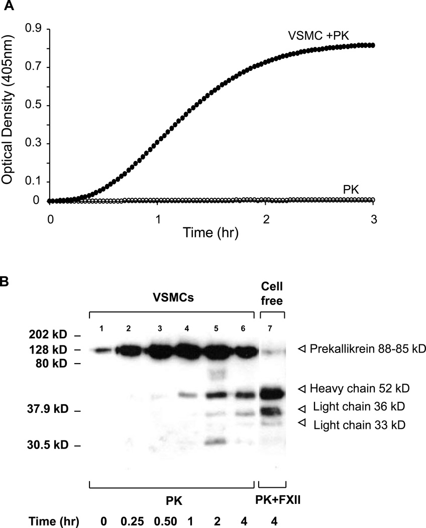
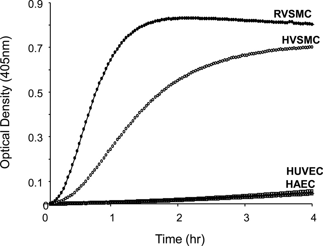
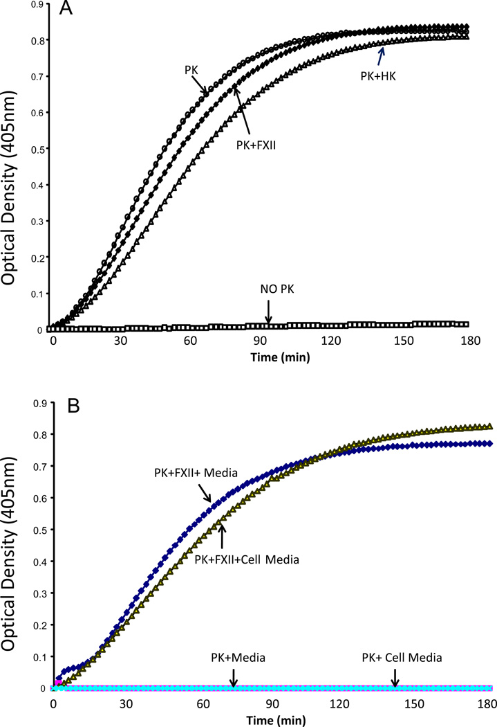
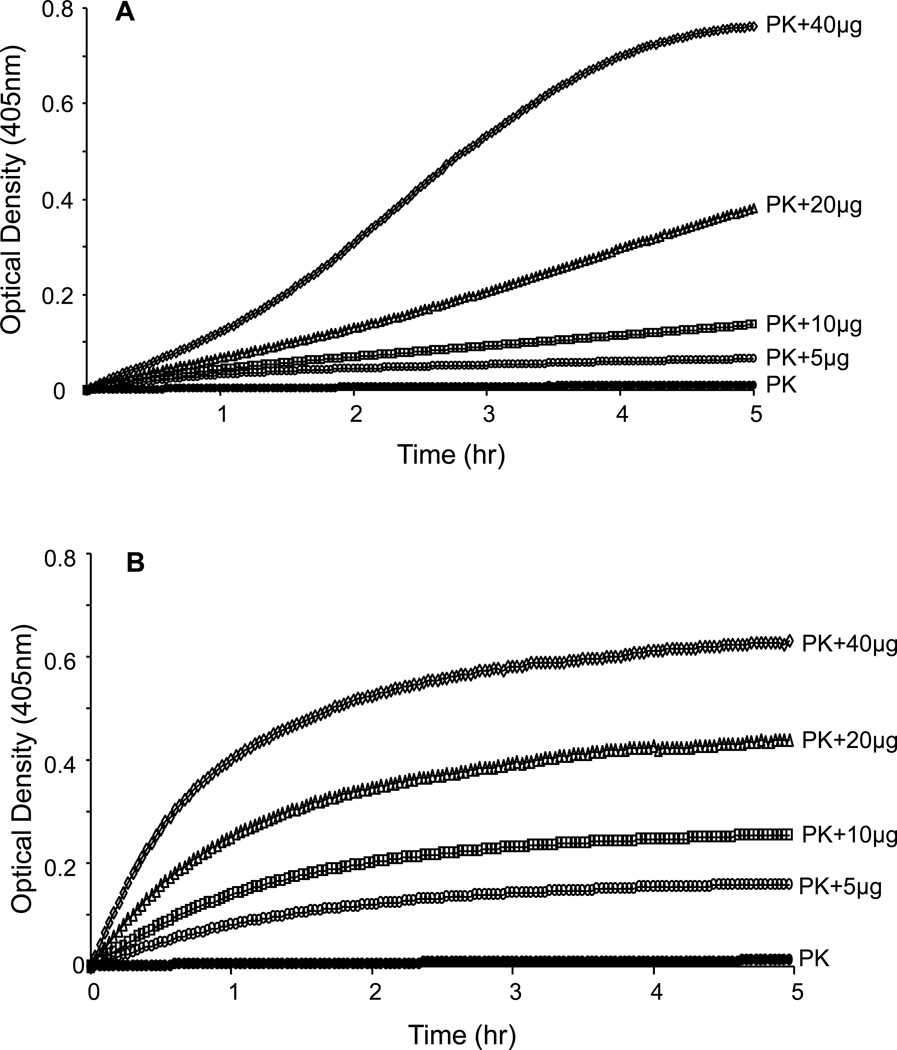
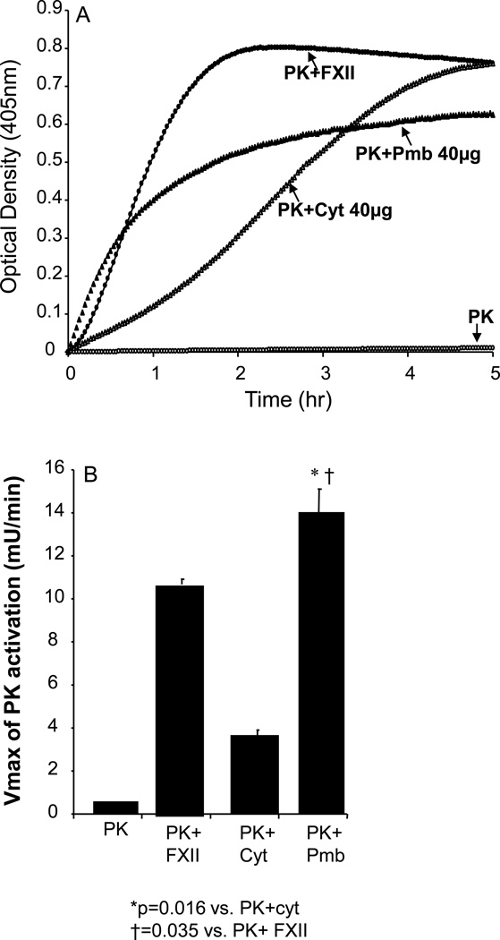
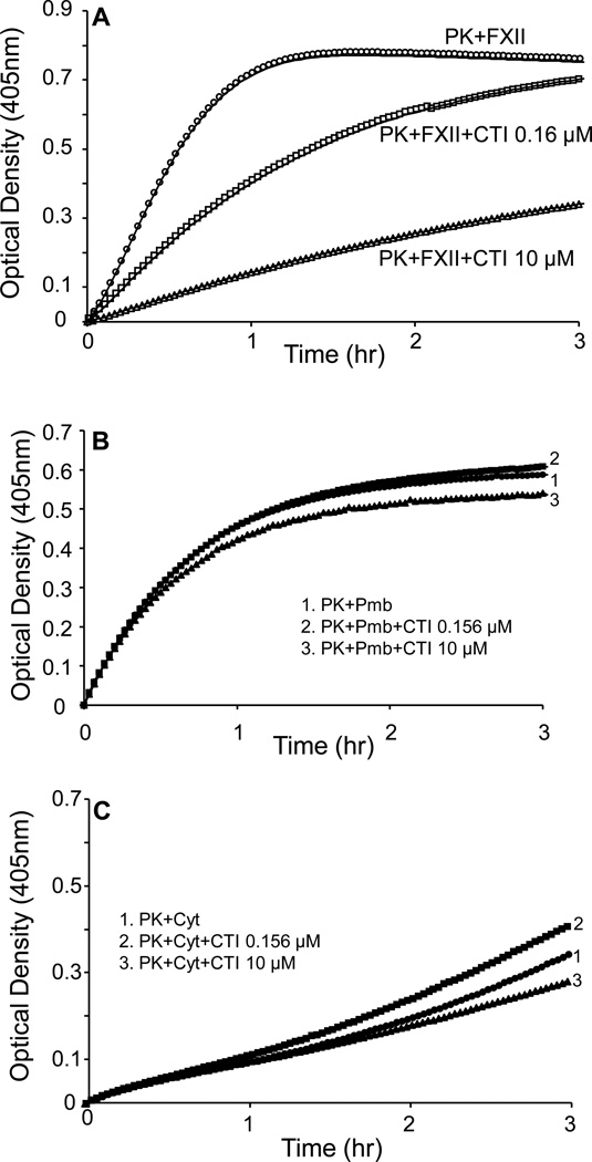
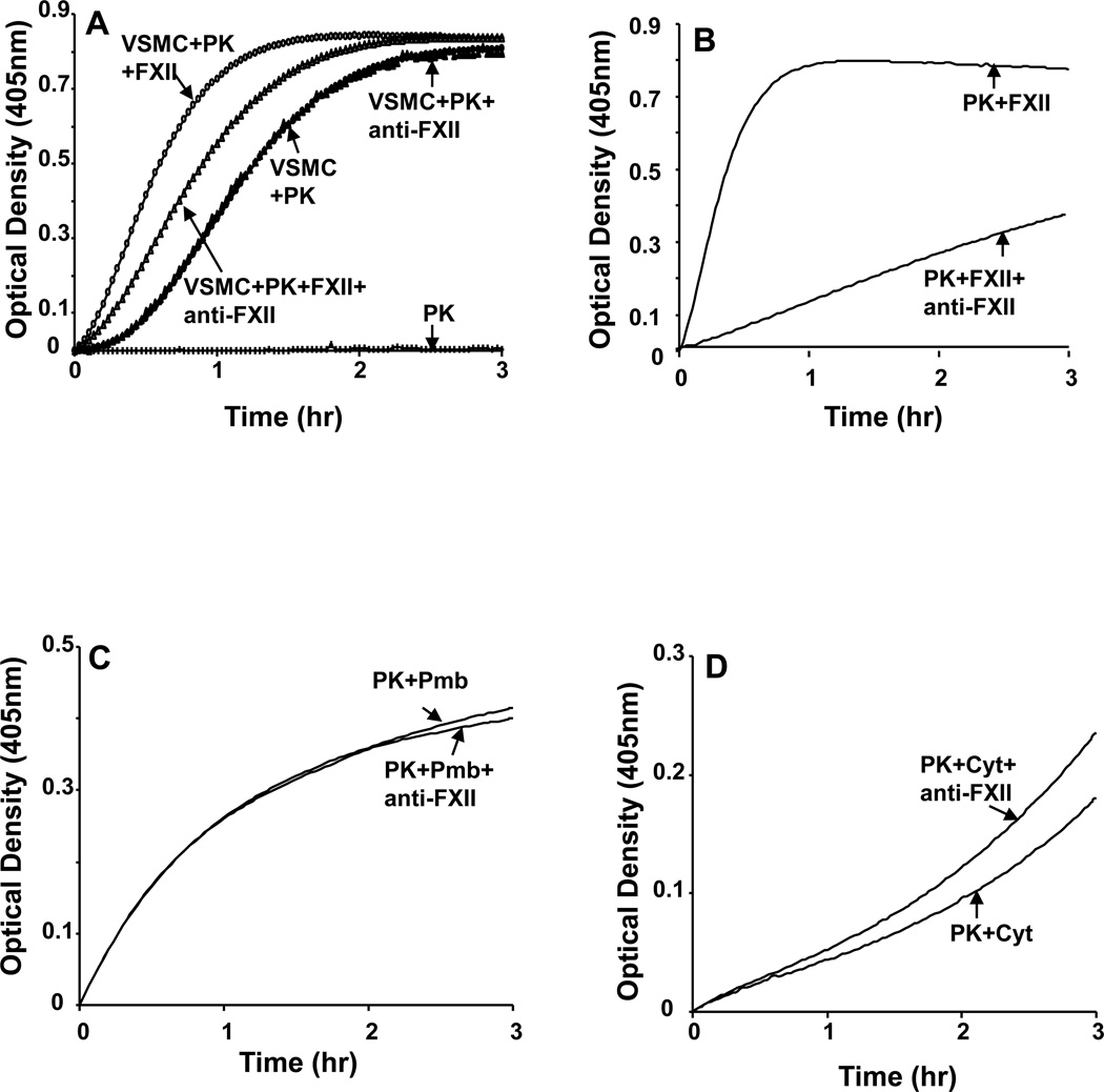
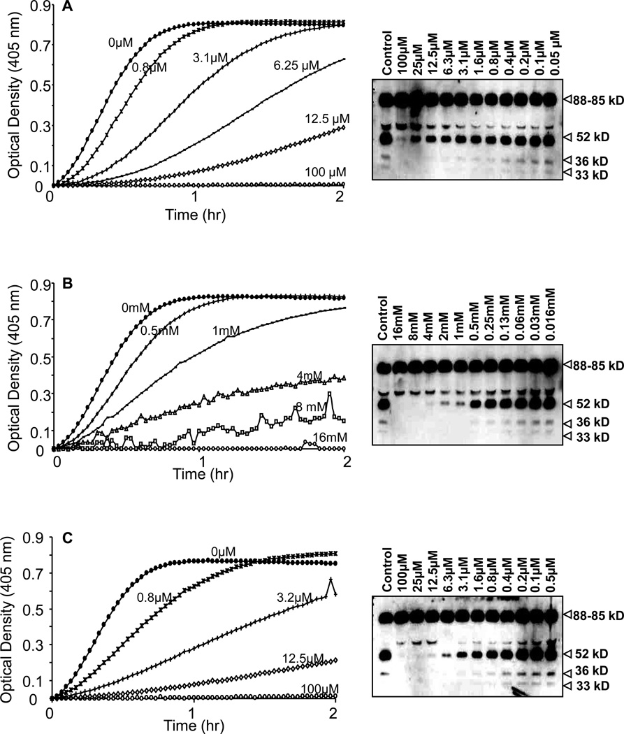
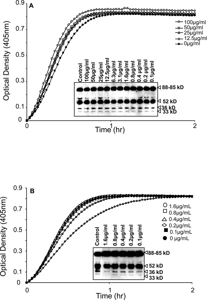
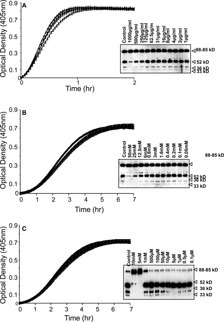
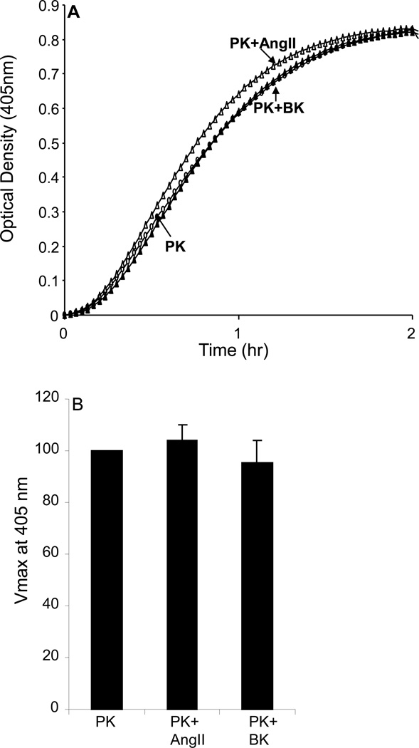
The contribution of plasma prekallikrein (PK) to vascular remodeling is becoming increasingly recognized. Plasma PK is activated when the zymogen PK is digested to an active enzyme by activated factor XII (FXII). Here, we present our findings that vascular smooth muscle cells (VSMC) activate plasma PK in the absence of FXII. Extracted plasma membrane and cytosolic fractions of VSMCs activate PK, but the rate of PK activation was greater by the membrane fraction. FXII neutralizing antibody did not affect PK activation by extracted proteins of VSMCs. VSMC PKA was inhibited by the serine protease inhibitors such as aprotinin, phenylmethylsulfonyl fluoride, leupeptin and CTI with CI50 of 0.78 μM, 1 mM, 3.13 μM and 40 nM on the cultured cells, respectively. No inhibition of PK activation by cysteine, aspartic acid, and metalloprotease inhibitors was observed. This is the first report of the presence of an intrinsic PKA in VSMC. Considering that VSMCs are normally separated from the circulating blood by endothelial cells, direct PK activation by VSMCs may play a role in disease states like diabetes, hyperlipidemia or hypertension where the endothelial layer is damaged.


Figures

Figure 1
Figure 1. Prekallikrein activation by cultured VSMCs in the absence of FXII
A: VSMCs were subcultured in the 96-well microplate. Optical densities (OD) were recorded at 405 nm at 2-min intervals for 3 hrs using SpectraMax® 340 PC laser microplate reader. No activation of PK was present by using 3.85 nM of PK and 0.4 mM of kallikrein substrate S-2302® in the cell-free system (open circle). When the same amount of PK was added to VSMCs, activation of PK was detected in the absence of FXII (closed circle). The graphs are from a representatives set of 10 experiments. B, Immunoblot analysis of conditioning media of VSMCs incubated with 3.58 nM of prekallikrein shows the conversion of PK into various digestion fragments including those corresponding to heavy chain (52 kD) and light chains (36 and 33 kD) of the PK as a function of time. PK digestion fragments by FXII in cell-free system are shown for comparison.
Figure 2
Figure 2. Comparison of PK activation by endothelial and VSMC
Rat aortic VSMCs, human aortic VSMCs (HVSMCs), human umbilical vein endothelial cells (HUVECs) and human aortic endothelial cells (HAECs) were subcultured in different wells and incubated with 3.58 nM of PK and 0.4 mM of S-2302. Optical densities were recorded at 405 nm at every 2 min for 2 hrs. While HUVECs and HAECs did not show PK activation, rat VSMCs (closed circle) and HVSMCs (open circle) showed remarkable activation of PK in the absence of FXII. Graphs are from a representative set of 3 experiments.
Figure 3
Figure 3
(A) Cleavage of S2303 by PK in VSMC. Rat aortic VSMCs were incubated with 0.4 mM of S-2302 in the presence and absence of 3.58 nM of PK alone and in the presence of FXII and/or HK. Optical densities were recorded at 405 nm at every 2 min for 3 hrs. Cleavage of S2302 by VSMC was observed only in the presence of PK, PK+FXII and PK+HK. No cleavage of S2302 was observed by VSMC in the absence of PK (NO PK). Graphs are from a representative set of 4 experiments. (B). Effect of cell culture media on PK activation. 0.4 mM of S-2302 and 3.58 nM of PK were incubated with conditioned media collected 24 h after incubation with VSMC in the presence and absence of FXII. No hydrolysis of S2302 by PK was observed in conditioned media (PK+Cell Media) collected from VSMC and in PK+ Media. Hydrolysis of S2302 by PK was observed only in the presence of FXII. Graphs are from a representative set of 4 experiments.
Figure 4
Figure 4. Dose-response prekallikrein activation by cytosolic and plasma membrane fractions
A: Dose response of cytosolic fractions (5, 10, 20 and 40 µg) on PK activation (3.58 nM of PK, closed circles). B: Dose response of plasma membrane fraction (Pmb) on PK activation in a cell free system. The results are representative of 4 separate experiments.
Figure 5
Figure 5. Prekallikrein activation by extracted proteins from VSMCs in cell-free system
A: 40 µg of cytosolic (Cyt) and/or plasma membrane (Pmb) protein fractions extracted from VSMC were used to assess activation of PK in cell-free system. Optical densities were read at 405 nm at 2-min intervals for 5 hr. Autoactivation of PK was not observed in the absence of cellular protein (open circle). Both cytosolic fraction (open triangle) and plasma membrane fraction (closed triangle) activated PK. B: The rate of PK activation by plasma membrane fraction was faster than that by cytosolic protein based on the maximum rate of the kinetic profile (Vmax). The Vmax of plasma membrane was even higher than that by factor XII (FXII) (closed circle in A). The results are averages from 3 experiments, each with triplicates.
Figure 6
Figure 6. Inhibitory profile of PK activation BY CTI in cell-free system
A, PK activation by FXII in the absence of CTI is shown. Using 0.156 µM and 10 µM, CTI inhibited activation of PK by factor XII (FXII) in dose-dependent manner (p=0.0161 and p=0.000176 for 0.156 µM and 10 µM, respectively, n=3). B, 40 µg of plasma membrane fraction (Pmb) of VSMC activated PK was not inhibited by 0.156 µM and 10 µM of CTI (p=0.996 and p=0.906 for 0.156 µM and 10 µM, respectively, n=3). C, Cytosolic fraction also activated PK, which did not show a significant inhibition by 0.156 µM and 10 µM of CTI (p=0.165 and p=0.765 for 0.156 µM and 10 µM, respectively, n=3).
Figure 7
Figure 7. The effect of anti-FXII antibody on the activation of PK
A: VSMCs incubated with anti-FXII antibody for 30 min at room temperature. Anti-FXII (100µg/ml) antibody inhibited PK activation by FXII (p=0.00674). However, activation of PK by the VSMCs in the absence of FXII was not inhibited by 100µg/mL of anti-FXII (closed circles and closed triangles). B, Anti-FXII inhibited PK activation by FXII (p=0.0144). C: Anti-FXII did not inhibit PK activation by plasma membrane fraction (Pmb, 40 µg) of VSMCs. D, Anti-FXII did not inhibit PK activation by cytosolic fraction (Cyt, 40µg) of VSMCs. The graphs are representative sets from 4 separate experiments.
Figure 8
Figure 8. Inhibition profile of PK activation on surfaces of VSMCs by serine protease inhibitors
A–C, PK activation by VSMCs was dose-dependently inhibited by aprotinin (A), phenylmethylsulfonyl fluoride (PMSF) (B) and leupeptin (C). Each graph of PK activation and the bands obtained by Western blotting represent the effect of serial dilution of protease inhibitors used (n=3).
Figure 9
Figure 9. Inhibitory profile of PK activation by cysteine and aspartic acid protease inhibitors
A, PK activation by VSMCs was not inhibited by cystatin, a cysteine protease inhibitor. B, PK activation by VSMCs was not inhibited by pepstatin, an aspartic acid protease inhibitor (n=3 experiments).
Figure 10
Figure 10. Inhibition profile of PK activation by metalloprotease inhibitors
A–C, PK activation by VSMCs was not inhibited by bestatin (A), EDTA-Na2 (B) and EGTA (C). Activation assays and western blot analysis showed similar results (n=3 experiments).
Figure 11
Figure 11. Effect of angiotensin II (AngII) and bradykinin (BK) on prekallikrein (PK) activation by VSMCs
PK activation was done in the absence (open circle) or presence of AngII (10 µM, open triangle) and BK (100 µM, closed triangle) for 2 hrs at room temperature. No inhibition of PK activation by VSMCs was observed by incubating PK in the presence of AngII or BK. The graphs were representative of 3 separate experiments.

Similar articles

Cited by

References

    1. Ross R. Atherosclerosis: An inflammatory disease. NEJM. 1999;340:115–126. - PubMed
    1. Clowes AW, Karnovsky MJ. Suppression by heparin of smooth muscle cell proliferation in injured arteries. Nature. 1977;265:625–626. - PubMed
    1. Jackson CL, Schwartz SM. Pharmacology of smooth muscle cell replication. Hypertension. 1992;20:713–736. - PubMed
    1. Luscher TF, Tanner FC, Tschudi MR, Noll G. Endothelial dysfunction in coronary artery disease. Annu Rev Med. 1993;44:395–418. - PubMed
    1. Shaw Jl, Diamandis EP. Distribution of 15 human kallikreins in tissues and biological fluids. Clin Chem. 2007;53:1423–1432. - PubMed

Publication types

LinkOut - more resources