. 2015 Jan 9;8(1):1.
doi: 10.1186/1756-8935-8-1. eCollection 2015.

Selective impairment of methylation maintenance is the major cause of DNA methylation reprogramming in the early embryo

Affiliations

Selective impairment of methylation maintenance is the major cause of DNA methylation reprogramming in the early embryo

Julia Arand et al. Epigenetics Chromatin. .

Abstract

Background: DNA methylomes are extensively reprogrammed during mouse pre-implantation and early germ cell development. The main feature of this reprogramming is a genome-wide decrease in 5-methylcytosine (5mC). Standard high-resolution single-stranded bisulfite sequencing techniques do not allow discrimination of the underlying passive (replication-dependent) or active enzymatic mechanisms of 5mC loss. We approached this problem by generating high-resolution deep hairpin bisulfite sequencing (DHBS) maps, allowing us to follow the patterns of symmetric DNA methylation at CpGs dyads on both DNA strands over single replications.

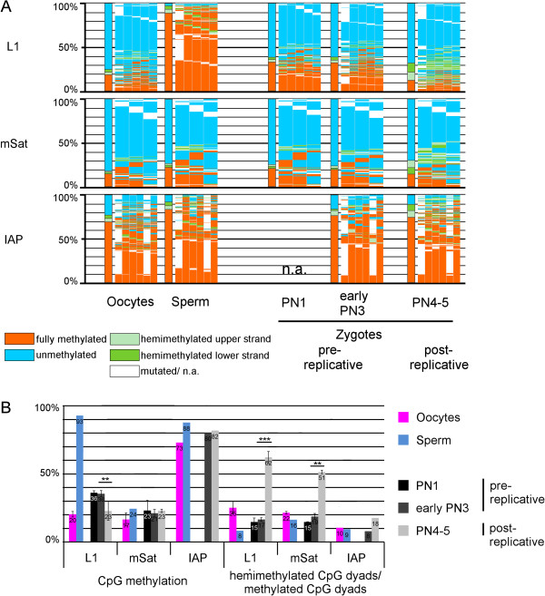
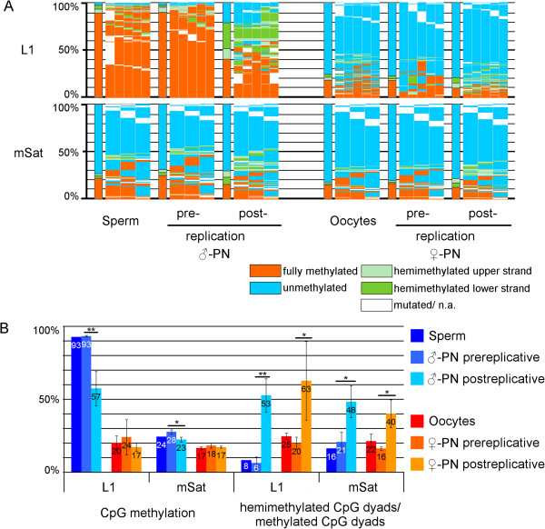
Results: We compared DHBS maps of repetitive elements in the developing zygote, the early embryo, and primordial germ cells (PGCs) at defined stages of development. In the zygote, we observed distinct effects in paternal and maternal chromosomes. A significant loss of paternal DNA methylation was linked to replication and to an increase in continuous and dispersed hemimethylated CpG dyad patterns. Overall methylation levels at maternal copies remained largely unchanged, but showed an increased level of dispersed hemi-methylated CpG dyads. After the first cell cycle, the combined DHBS patterns of paternal and maternal chromosomes remained unchanged over the next three cell divisions. By contrast, in PGCs the DNA demethylation process was continuous, as seen by a consistent decrease in fully methylated CpG dyads over consecutive cell divisions.

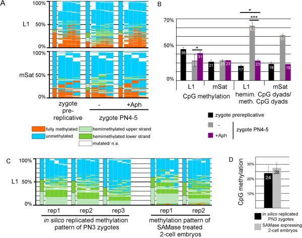
Conclusions: The main driver of DNA demethylation in germ cells and in the zygote is partial impairment of maintenance of symmetric DNA methylation at CpG dyads. In the embryo, this passive demethylation is restricted to the first cell division, whereas it continues over several cell divisions in germ cells. The dispersed patterns of CpG dyads in the early-cleavage embryo suggest a continuous partial (and to a low extent active) loss of methylation apparently compensated for by selective de novo methylation. We conclude that a combination of passive and active demethylation events counteracted by de novo methylation are involved in the distinct reprogramming dynamics of DNA methylomes in the zygote, the early embryo, and PGCs.

Keywords: DNA methylation pattern; DNA methylation reprogramming; Deep hairpin bisulfite sequencing; Pre-implantation development.


Figures

Figure 1
Figure 1
DNA Methylation patterns of L1Md_Tf (L1), major satellites (mSat), and IAPLTR1 (IAP) in germ cells and three pronuclear stages (PN) of mouse zygotes. (A) DNA methylation patterns, Bars are the sum of the DNA methylation status of all CpG dyads. The map next to the bar represents the distribution of methylated sites. Each column shows neighbored CpG dyads, and each line represents one sequence read. The reads in the map are sorted first by fully methylated sites and then by hemimethylated CpG dyads. Red, fully methylated CpG dyads; light green and dark green, hemimethylated CpG dyads on the upper and lower strand; blue, unmethylated CpG dyads; white, mutated or not analyzable. As 5-methylcytosine (5mC) and 5-hydroxymethylcytosine (5hmC) cannot be discriminated by bisulfite sequencing, "mC" should be considered to mean 5mC or 5hmC throughout the paper, and equally "C" (cytosine) should be considered to mean C, 5-formylcytosine (5fC), or 5-carboxycytosine (5caC). (B) Absolute DNA methylation level and percentage of hemimethylated CpG dyads in relation to all methylated CpG dyads. DNA Methylation patterns of L1, mSat and IAP were analyzed in germ cells (oocytes and sperm) and different PN stages (PN1 and early PN3 are before replication, PN4 to PN5 are after replication) using deep hairpin bisulfite sequencing (DHBS). DNA methylation pattern changes can be observed following the first DNA replication after fertilization; in all elements an increasing amount of hemimethylated CpG dyads can be seen, and for L1 DNA demethylation can also be observed.
Figure 2
Figure 2
DNA methylation patterns of major satellites (mSat) and L1Md_Tf (L1) of separated pronuclei of pre- and post-replicative stages of mouse zygotes and germ cells. (A) DNA methylation patterns; for explanation, see Figure  1, (B) Absolute DNA methylation level and percentage of hemimethylated CpG dyads in relation to all methylated CpG dyads. Paternal and maternal pronuclei were separated by micromanipulation before and after DNA replication, and L1 and mSat were analyzed with deep hairpin bisulfite sequencing (DHBS). As comparison, we added DHBS data from germ cells. Only after replication was a decrease in DNA methylation found on paternal chromosomes; accompanied by an increase in hemimethylated CpG dyads on both maternal and paternal chromosomes.
Figure 3
Figure 3
Replication dependency of DNA methylation reprogramming in the zygote. (A) DNA methylation patterns (for explanation see Figure  1) and (B) absolute DNA methylation level and percentage of hemimethylated CpG dyads in relation to all methylated CpG dyads of L1 and mSat in +/- aphidicolin-treated (+/- replication-blocked) PN4-5 zygotes. Aphidicolin treatment leads to diminished DNA demethylation. (C,D) DNA methylation patterns of replicates (rep) and (C) absolute DNA methylation level of SAMase-treated early (pre-replicative) two-cell embryos. SAMase diminishes all methylation events that are dependent on S-adenosyl-methionine (SAM). The patterns were compared with those of in silico replicated zygotes with no DNA methylation maintenance (see Methods). The DNA methylation profiles of the biological replication without methylation events (SAMase-treated two-cell embryos) are very similar to those events simulated in silico without methylation.
Figure 4
Figure 4
DNA methylation pattern of L1Md_Tf (L1), major satellites (mSat), and IAPLTR1 (IAP) in cleavage stage mouse embryos. (A) DNA methylation patterns (for explanation see Figure  1) (B) Absolute DNA methylation levels and percentage of hemimethylated CpG dyads in relation to all methylated CpG dyads. Two-cell embryos were analyzed before and after replication, and further-cleavage stage embryos were analyzed at 12-hour intervals to determine methylation changes during replications. DNA methylation pattern remained stable until the morula stage, when a further drop in methylation occurred at L1 and IAP. Interestingly, over the course of the cleavage stages the amount of hemimethylated CpG positions remained equally high.
Figure 5
Figure 5
DNA Methylation pattern of L1Md_Tf (L1), major satellites (mSat), and IAPLTR1 (IAP) in primordial germ cells (PGCs). (A) DNA methylation patterns (for explanation see Figure  1). (B) Absolute DNA methylation levels and percentage of hemimethylated CpG dyads in relation to all methylated CpG dyads. PGCs were sorted and analyzed at 24-hour intervals from 9.5 until 13.5 days post-coitum (dpc). DNA methylation decreased continuously, with a stable relative level of hemimethylated CpG positions.
Figure 6
Figure 6
Summary of DNA methylation changes in primordial germ cells (PGCs) and pre-implantation embryos. (A) Absolute DNA methylation levels and (B) percentage of hemimethylated CpG dyads in relation to all methylated CpG dydas (red, L1Md_Tf (L1); blue, major satellites (mSat); green, IAPLTR1 (IAP)). Note that from 13.5 dpc PGCs to the two-cell stage the values are depicted separately for maternal and paternal chromosomes, respectively, and thereafter depicted as combined values.

References

    1. Reik W, Dean W, Walter J. Epigenetic reprogramming in mammalian development. Science. 2001;293:1089–1093. doi: 10.1126/science.1063443. - DOI - PubMed
    1. Popp C, Dean W, Feng S, Cokus SJ, Andrews S, Pellegrini M, Jacobsen SE, Reik W. Genome-wide erasure of DNA methylation in mouse primordial germ cells is affected by AID deficiency. Nature. 2010;463(7284):1101–1105. doi: 10.1038/nature08829. - DOI - PMC - PubMed
    1. Smallwood SA, Tomizawa S, Krueger F, Ruf N, Carli N, Segonds-Pichon A, Sato S, Hata K, Andrews SR, Kelsey G. Dynamic CpG island methylation landscape in oocytes and preimplantation embryos. Nat Genet. 2011;43(8):811–814. doi: 10.1038/ng.864. - DOI - PMC - PubMed
    1. Guibert S, Forne T, Weber M. Global profiling of DNA methylation erasure in mouse primordial germ cells. Genome Res. 2012;22(4):633–641. doi: 10.1101/gr.130997.111. - DOI - PMC - PubMed
    1. Seisenberger S, Andrews S, Krueger F, Arand J, Walter J, Santos F, Popp C, Thienpont B, Dean W, Reik W. The dynamics of genome-wide DNA methylation reprogramming in mouse primordial germ cells. Mol Cell. 2012;48(6):849–862. doi: 10.1016/j.molcel.2012.11.001. - DOI - PMC - PubMed

LinkOut - more resources