Review
. 2015 Jan 18;6(1):117-26.
doi: 10.5312/wjo.v6.i1.117.

Current strategies for the restoration of adequate lordosis during lumbar fusion

Affiliations
Review

Current strategies for the restoration of adequate lordosis during lumbar fusion

Cédric Barrey et al. World J Orthop. .

Abstract

Not restoring the adequate lumbar lordosis during lumbar fusion surgery may result in mechanical low back pain, sagittal unbalance and adjacent segment degeneration. The objective of this work is to describe the current strategies and concepts for restoration of adequate lordosis during fusion surgery. Theoretical lordosis can be evaluated from the measurement of the pelvic incidence and from the analysis of spatial organization of the lumbar spine with 2/3 of the lordosis given by the L4-S1 segment and 85% by the L3-S1 segment. Technical aspects involve patient positioning on the operating table, release maneuvers, type of instrumentation used (rod, screw-rod connection, interbody cages), surgical sequence and the overall surgical strategy. Spinal osteotomies may be required in case of fixed kyphotic spine. AP combined surgery is particularly efficient in restoring lordosis at L5-S1 level and should be recommended. Finally, not one but several strategies may be used to achieve the need for restoration of adequate lordosis during fusion surgery.

Keywords: Lumbar lordosis; Pelvis incidence; Pelvis shape; Sagittal balance; Spinal fusion; Spine surgery.


Figures

Figure 1
Figure 1
Hypolordotic lumbo-sacral fusion with hyperextension of the segment above the instrumentation. Failure to restore a good sagittal balance leads to chronic back pain and early degenerative changes at adjacent level(s).
Figure 2
Figure 2
Duval-Beaupère’s pelvic parameters. Sacral slope (SS), pelvic tilt (PT), pelvic incidence (PI) and mathematical relation between the parameters (PI = SS + PT).
Figure 3
Figure 3
Low pelvic incidence is usually associated with slight sacral slope and flat lumbar spine, and high pelvic incidence with great sacral slope and more curved lumbar spine[32]. PI: Pelvis incidence; PT: Pelvis tilt.
Figure 4
Figure 4
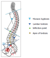
View of several spinal parameters. Lumbar lordosis (LL), Thoracic kyphosis (TK), Apex of the lordosis, Inflection point. PI: Pelvis incidence; PT: Pelvis tilt; SS: Sacral slope.
Figure 5
Figure 5
Roussouly’s classification of sagittal profiles of the spine in four types[33].
Figure 6
Figure 6
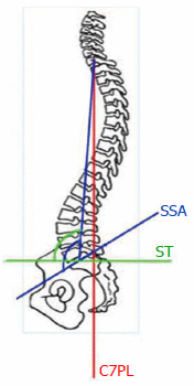
C7 plumb line, spino sacral angle and spinal tilt. C7PL: C7 Plumb Line; SSA: Spino sacral angle; ST: Spinal tilt.
Figure 7
Figure 7
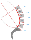
Sagittal lumbar curvature modeled by a portion of an ellipse. Note that 2/3 of the lumbar lordosis is located in the lower lumbar spine between L4 and S1 and that 85% of the lordosis is given by the L3-S1 segment.
Figure 8
Figure 8
Different positions on operative table (Figures on the left coming from the work by Stephens et al[37]. Clinical case on the right: Loss of lumbar lordosis in knee-chest position with L2-S1 angle passing from 50° preoperatvely to 28° peroperatively).
Figure 9
Figure 9
The importance of rod contouring depends on the spino-pelvic morphotype. PI: Pelvic incidence.
Figure 10
Figure 10
Impact of rod-screw connection on lordosis restoration: monoaxial vs polyaxial screws. With mono-axial screws, the connection between the screw and the rod is perpendicular, permitting therefore to position the spine approximately in a similar position compared to the rod. On the contrary, using poly-axial screws, the connection between the screw and the rod is angulated with the spine less lordotic compared to the rod. In the figure, although the contouring of the rod is similar for the two spines, the spine on the right, instrumented with poly-axial screws, is significantly less lordotic.
Figure 11
Figure 11
Posterior and lateral view of combined anterior/posterior arthrodesis using anterior lumbar interbody fusion cage inserted anteriorly and posterior pedicle-screw stabilization[55].

References

    1. Doherty J. Complications of fusion in lumbar scoliosis. Proc Scoliosis Res Soc. 1973;55:438–45.
    1. Moe JH, Denis F. The iatrogenic loss of lumbar lordosis. Orthop Trans. 1977;1:131.
    1. Swank S, Lonstein JE, Moe JH, Winter RB, Bradford DS. Surgical treatment of adult scoliosis. A review of two hundred and twenty-two cases. J Bone Joint Surg Am. 1981;63:268–287. - PubMed
    1. Swank SM, Mauri TM, Brown JC. The lumbar lordosis below Harrington instrumentation for scoliosis. Spine (Phila Pa 1976) 1990;15:181–186. - PubMed
    1. Lagrone MO, Bradford DS, Moe JH, Lonstein JE, Winter RB, Ogilvie JW. Treatment of symptomatic flatback after spinal fusion. J Bone Joint Surg Am. 1988;70:569–580. - PubMed