. 2015 Jan 9:2:74.
doi: 10.3389/fcell.2014.00074. eCollection 2014.

Polyethylenimine-mediated expression of transgenes in the acinar cells of rats salivary glands in vivo

Affiliations

Polyethylenimine-mediated expression of transgenes in the acinar cells of rats salivary glands in vivo

Monika Sramkova et al. Front Cell Dev Biol. .

Abstract

Non viral-mediated transfection of plasmid DNA provides a fast and reliable way to express various transgenes in selected cell populations in live animals. Here, we show an improvement of a previously published method that is based on injecting plasmid DNA into the ductal system of the salivary glands in live rats. Specifically, using complexes between plasmid DNA and polyethyleneimine (PEI) we show that the expression of the transgenes is directed selectively to the salivary acinar cells. PEI does not affect the ability of cells to undergo regulated exocytosis, which was one of the main drawbacks of the previous methods. Moreover PEI does not affect the proper localization and targeting of transfected proteins, as shown for the apical plasma membrane water channel aquaporin 5 (AQP5). Overall, this approach, coupled with the use of intravital microscopy, permits to conduct localization and functional studies under physiological conditions, in a rapid, reliable, and affordable fashion.

Keywords: acinar cells; aquaporin 5; in vivo transfection; intravital microscopy; non-viral gene delivery; polyethyleneimine; salivary glands.


Figures
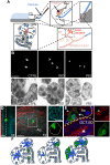
Figure 1
Figure 1
Non viral-mediated expression of transgenes in rat salivary glands. (A) Diagram of the transfection procedures in anesthetized rats. A thin polyethylene cannula is inserted into the Wharton's duct, as described in the Methods sections. The plasmid DNA is gently injected and diffuses into the large ducts (granular convoluted tubules or GCT, and the striated ducts or SD), then into the intercalated ducts (IC), and finally into the acinar canaliculi. Plasmid DNA accesses the epithelium through the apical plasma membrane (APM, arrows). (B) In vivo transfection with plasmid DNA -12 μg of Plasmid DNA encoding for pVenus were injected alone (CTRL, left panel), after SC injection of isoproterenol (ISO, center panel), or mixed with PEI as described in the Methods (PEI, right panel). After 16 h the submandibular SGs were excised and immediately imaged by two-photon microscopy using a 10X objective (excitation 930 nm) to reveal the cells expressing pVenus (arrows). Scale bar, 1 mm. (C) Effect of PEI on regulated exocytosis. Mice expressing cytoplasmic GFP were treated as described in B and the SGs imaged by confocal microscopy (excitation 488 nm) to estimate exocytosis of the secretory granules, as previously reported (Milberg et al., 2013). In the inset the apical plasma membrane (arrows) and the secretory granules (arrowheads) are highlighted. Scale bar, 20 μm. (D–E) PEI-mediated transfection in SGs drives expression into acinar cells. (D) Excised glands transfected with pVenus/PEI were imaged by two-photon microscopy using a 60x water immersion objective (D) or processed for immunofluorescence and imaged by confocal microscopy (E). (D) Two Z-stacks from the same area of the sample were acquired twice by using two different excitation wavelengths, respectively: 740 nm to reveal the epithelium of the gland (cyan) and 930 nm to reveal pVenus (green) and collagen fibers (red). The Z-stacks were combined and shown as xz (left) or xy (center) projection. The cell expressing pVenus is part of an acinus (inset, right). Scale bar, 20 μm. (E) The SGs were labeled for phallodin (red) and the nuclear staining Hoechst (blue). The cell expressing pVenus (green) is part of an acinus (white broken line) as identified by the actin-labeled acinar canaliculi (arrowhead). Large ducts (yellow broken line), as identified by the characteristic actin pattern (asterisk) did not express any pVenus. Scale bar, 10 μm. (F) Diagram summarizing the pattern of expression of plasmid DNA in SGs. pVenus was expressed in cells of the IC under control conditions (left) and in acinar cells either upon stimulation with ISO (center) or administration with PEI (right).
Figure 2
Figure 2
PEI enables the expression of several transgenes in the acinar cells of the SGs. (A–C) Plasmid DNAs encoding either for selected fluorescently tagged proteins (A,B) or for multiple copies of pVenus (C) were injected into the SGs of anesthetized rats under different conditions. (A) High magnification images acquired by two-photon microscopy (excitation 930 nm, 60x water-immersion objective) to reveal the intracellular localization of selected transfected transgenes. (B–C) The efficiency of expression of the transgene was determined as described in the Methods and expressed as cells/mm2. Values represent average ±S.D. (N = 3). Statistical significance was calculated by using a t-test *p < 0.1 ***p < 0.001 ****P < 0.0001. (B) Plasmids encoding for various cellular proteins and mixed with PEI are shown. (C) Plasmids containing 1 (1X), 2 (2X) or 3 (3X) copies of pVenus fused together where injected into the salivary duct alone, after stimulation with ISO, or mixed with PEI. (D) Plasmid DNA encoding for the F-actin reporter RFP-lifeact was mixed with PEI and injected into the salivary duct of anesthetized rats. After 24 h the SGs were exposed and imaged by confocal microscopy (excitation 561 nm). In acinar cells, RFP-life act was loclaized at the plasma membrane and in particular at the APM (arrows). SC injection of isoproterenol (time 0:00) elicited regulated exocytosis. As previously shown (Masedunskas et al., 2011a), secretory granules fused with the APM and recruited a F-actin coat (arrowheads). Scale bar, 10 μm.
Figure 3
Figure 3
Characterization of the fluorescently tagged AQP5. (A) Human AQP5 was tagged either at the (C) or the N-terminus and transiently expressed in HSG cells grown on glass slides. AQP5-YFP is localized in the ER and the nuclear envelope (arrows), whereas YFP-AQP5 is localized at the APM (arrowheads). (B–D) HSG cells (B, C, and D lower panel) or HSG cells stably expressing YFP-AQP5 (D, upper panel) were grown for 96 h in matrigel to form acinar-like structures or on glass coverslips (C, upper panel) and imaged by either phase contrast (B, upper panel) or confocal microscopy (B, C, lower panel and C) in order to acquire Z-stacks. Cells were fixed and processed to reveal F-actin and the nuclei (B, lower panel), YFP-AQP5 (C, upper panel), endogenous AQP5 (C, lower panel), amylase (C) and the Golgi apparatus (GM130, C). Maximal projections of the xy view and side views (xz, yz) show that F-actin is partially polarized in the acinar-like cells (arrows) whereas both endogenous and ectopically expressed AQP5 did not. Scale bars, 5 μm.
Figure 4
Figure 4
Targeting of ectopically expressed AQP5 in the SG in vivo. The SGs of anesthetized rats were left untreated (D) or injected with a plasmid encoding for rat YFP-AQP5 either alone (A,B) or in combination with TGN38-mcherry, a marker for the Trans-Golgi Network (C,E). The salivary glands were exposed and imaged by two-photon microscopy (A,B,E) or fixed, processed for immunofluorescence and imaged by confocal microscopy (C,D). (A) Two Z-stacks from the same area of the sample were acquired twice by using two different excitation wavelengths, respectively: 740 nm to reveal the epithelium of the gland (cyan) and 930 nm to reveal the YFP (green) and collagen fibers (red). The Z-stacks were combined as described in the Methods and shown as maximal projections. Upper panel shows a low magnification of the parenchyma (20X objective) with 5 cells expressing YFP-AQP5 (arrows). Lower panel show an acinus (broken line) with a transfected cell (60X objective). Scale bars, 40 μm. (B) Maximal projection of an individual acinar cells (Left panel) and high magnification of a single optical slice (60X objective) showing that YFP-AQP5 (green) is localized a at the APM and in small vesicles (arrows). Scale bars, 5 μm. (C,D) Cryosections from SGs were labeled with Rhodamine-Phalloidin (red) alone (C), or with an antibody directed against rat AQP5 (D, green). Both ectopically expressed and endogenous AQP5 are localized at the APM (arrowheads) and in small vesicles (arrows). Scale bars, 5 μm. (E) Time lapse imaging of an individual acinar cells show that YFP-AQP5-containing vesicles are stationary over a long period of time. Note a TGN38-containng vesicle that pinches off the TGN (arrowhead). Scale bar 3 μm.

Similar articles

Cited by

References

    1. Adriaansen J., Zheng C., Perez P., Baum B. J. (2010). Production and sorting of transgenic, modified human parathyroid hormone in vivo in rat salivary glands. Biochem. Biophys. Res. Commun. 391, 768–772. 10.1016/j.bbrc.2009.11.135 - DOI - PMC - PubMed
    1. Amornphimoltham P., Rechache K., Thompson J., Masedunskas A., Leelahavanichkul K., Patel V., et al. . (2013). Rab25 regulates invasion and metastasis in head and neck cancer. Clin. Cancer Res. 19, 1375–1388. 10.1158/1078-0432.CCR-12-2858 - DOI - PMC - PubMed
    1. Boulaiz H., Marchal J. A., Prados J., Melguizo C., Aranega A. (2005). Non-viral and viral vectors for gene therapy. Cell. Mol. Biol. (Noisy-le-grand). 51, 3–22. - PubMed
    1. Creusat G., Rinaldi A. S., Weiss E., Elbaghdadi R., Remy J. S., Mulherkar R., et al. . (2010). Proton sponge trick for pH-sensitive disassembly of polyethylenimine-based siRNA delivery systems. Bioconjug. Chem. 21, 994–1002. 10.1021/bc100010k - DOI - PubMed
    1. Delporte C., Steinfeld S. (2006). Distribution and roles of aquaporins in salivary glands. Biochim. Biophys. Acta 1758, 1061–1070. 10.1016/j.bbamem.2006.01.022 - DOI - PubMed

LinkOut - more resources