

Case Reports
doi: 10.3904/kjim.1989.4.1.96.
A case of subcutaneous seeding of hepatocellular carcinoma after fine needle aspiration biopsy
- PMID: 2562134
- PMCID: PMC4534970
- DOI: 10.3904/kjim.1989.4.1.96
Item in Clipboard
Case Reports
A case of subcutaneous seeding of hepatocellular carcinoma after fine needle aspiration biopsy
Korean J Intern Med.
1989 Jan.
Abstract
Cancer spread along the needle track following fine needle aspiration biopsy is said to be a rare complication. The authors report a case of subcutaneous implantation of hepatocellular carcinoma following ultrasono-guided fine needle aspiration biopsy. The patient, a 67-year-old Korean male was found to have a large hepatocellular carcinoma diagnosed by fine needle aspiration biopsy. Four months later, the patient felt two subcutaneous growing lumps at the previous aspiration site. The authors confirmed them histologically 11 months after aspiration.
Figures
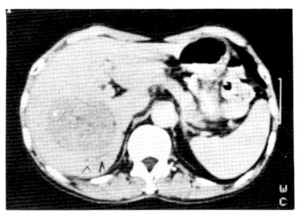
Abdominal CT scan showed an inhomogeneously enhanced hepatic tumor with well demarcation. (9 × 9 × 8 cm).
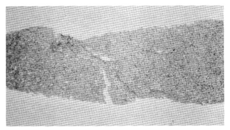
Chronic persistent hepatitis showing chronic inflammatory cell infiltration and widening of the portal tract.
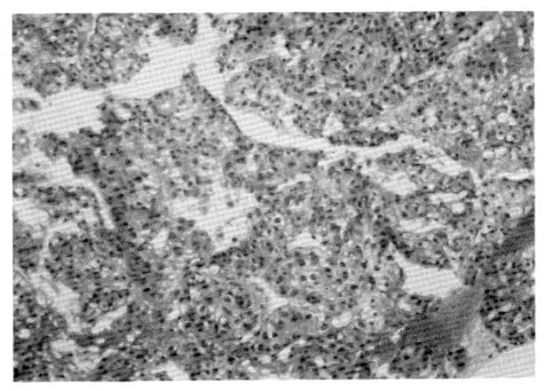
Hepatocellular carcinoma showing trabecular pattern with sinusoidal structure and pleomorphic epithelial cells resembling hepatocytes.
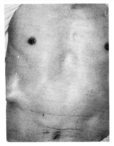
The subcutaneous lumps were noted at the previous ultrasonoguided needle biopsy site.
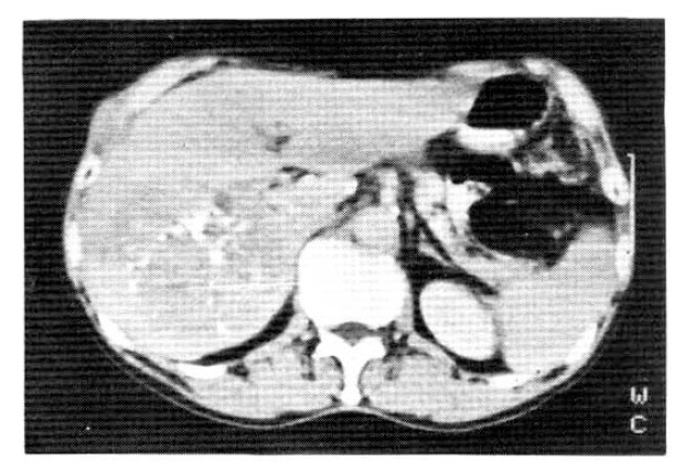
Abdominal CT scan showed a more enlarged size (10 × 10 × 11 cm) with evidence of metastasis to the abdominal wall and only a small amount of lipiodol droplets remaining in the tumor.
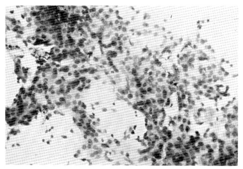
Irregular sheet of malignant epithelial cells which have anisonucleosis. hyperchromatism and occasional distinct nucleoli.
Similar articles
-
Subcutaneous seeding of hepatocellular carcinoma after percutaneous needle biopsy.Gut. 1999 Oct;45(4):626-7. doi: 10.1136/gut.45.4.626. Gut. 1999. PMID: 10486377 Free PMC article. Review. No abstract available.
-
[Subcutaneous seeding of hepatocellular carcinoma after fine-needle percutaneous biopsy].Rev Esp Enferm Dig. 2007 Jun;99(6):354-7. doi: 10.4321/s1130-01082007000600010. Rev Esp Enferm Dig. 2007. PMID: 17883301 Spanish.
-
[A case report of cutaneous seeding of hepatocellular carcinoma following ultrasound guided fine needle aspiration biopsy].Nihon Shokakibyo Gakkai Zasshi. 1990 May;87(5):1253-7. Nihon Shokakibyo Gakkai Zasshi. 1990. PMID: 2166846 Japanese. No abstract available.
-
Subcutaneous seeding of small hepatocellular carcinoma after fine needle aspiration biopsy.J Gastroenterol Hepatol. 1993 Mar-Apr;8(2):195-8. doi: 10.1111/j.1440-1746.1993.tb01513.x. J Gastroenterol Hepatol. 1993. PMID: 8386023
-
Needle track seeding of primary and secondary liver carcinoma after percutaneous liver biopsy.HPB Surg. 1993;6(3):199-203; discussion 203-4. doi: 10.1155/1993/39539. HPB Surg. 1993. PMID: 8387809 Free PMC article. Review.
Cited by
-
Diagnosis of HCC.Dig Dis Sci. 1997 Oct;42(10):2033-4. doi: 10.1023/a:1018854031015. Dig Dis Sci. 1997. PMID: 9365131 No abstract available.
-
Subcutaneous seeding of hepatocellular carcinoma after percutaneous needle biopsy.Gut. 1999 Oct;45(4):626-7. doi: 10.1136/gut.45.4.626. Gut. 1999. PMID: 10486377 Free PMC article. Review. No abstract available.
References
-
- Kline TS, Neal HS. Needle aspiration biopsy; A pilot study. JAMA. 1973;224:1143–1146. - PubMed
-
- Kline TS, Neal HS. Needle aspiration biopsy; A critical appraisal. JAMA. 1978;239:36–39. - PubMed
-
- Ferruci JT, Witterberg JW, Margolies MN, Carney RW. Malignant seeding of the tract after thin-needle aspiration biopsy. Radiology. 1979;130:345–346. - PubMed
-
- Smith FP, Macdonald JS, Schein PS, Ornitz RD. Cutaneous seeding of pancreas cancer by skinny-needle aspiration. Arch Intern Med. 1980:140–855. - PubMed
-
- Moloo Z, Finley RJ, Turner-Smith L, Craig ID. Possible spread of bronchogenic carcinoma to the chest wall after a thoracic fine needle aspiration biopsy. Acta Cytol. 1985;29:167–169. - PubMed
Publication types
MeSH terms
LinkOut - more resources
Full Text Sources
Medical