. 2015;7(2):377-89.
doi: 10.1080/19420862.2015.1007816.

Improving target cell specificity using a novel monovalent bispecific IgG design

Affiliations

Improving target cell specificity using a novel monovalent bispecific IgG design

Yariv Mazor et al. MAbs. 2015.

Erratum in

  • Corrigendum.
    [No authors listed] [No authors listed] MAbs. 2016 May-Jun;8(4):839. doi: 10.1080/19420862.2016.1175161. MAbs. 2016. PMID: 27142173 Free PMC article. No abstract available.

Abstract

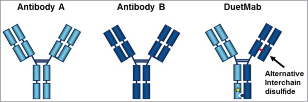
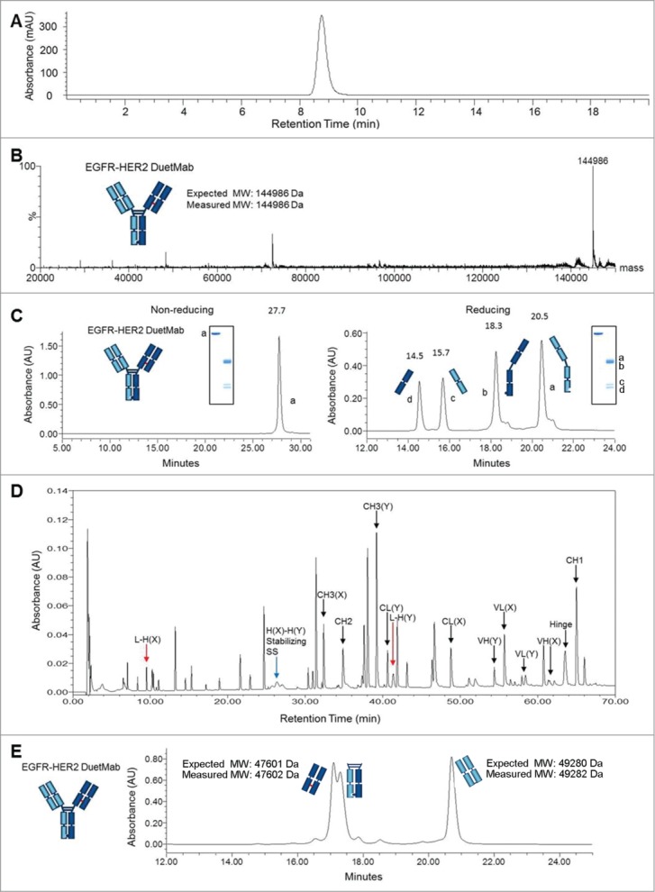
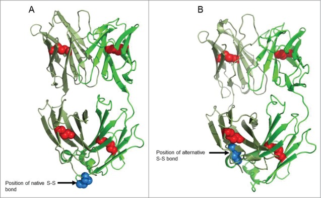
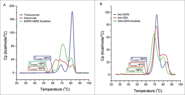
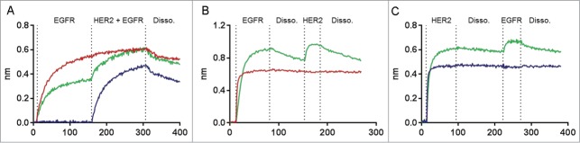
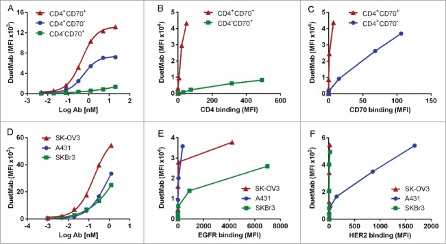
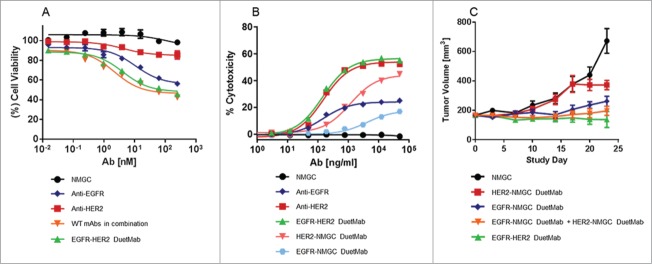
Monovalent bispecific IgGs cater to a distinct set of mechanisms of action but are difficult to engineer and manufacture because of complexities associated with correct heavy and light chain pairing. We have created a novel design, "DuetMab," for efficient production of these molecules. The platform uses knobs-into-holes (KIH) technology for heterodimerization of 2 distinct heavy chains and increases the efficiency of cognate heavy and light chain pairing by replacing the native disulfide bond in one of the CH1-CL interfaces with an engineered disulfide bond. Using two pairs of antibodies, cetuximab (anti-EGFR) and trastuzumab (anti-HER2), and anti-CD40 and anti-CD70 antibodies, we demonstrate that DuetMab antibodies can be produced in a highly purified and active form, and show for the first time that monovalent bispecific IgGs can concurrently bind both antigens on the same cell. This last property compensates for the loss of avidity brought about by monovalency and improves selectivity toward the target cell.

Keywords: ADCC, antibody-dependent cell-mediated cytotoxicity; Biotechnology; CDR, complementarity determining region; CH1, 2 and 3-heavy chain constant domain 1, 2 and 3; CL-, light chain constant domain; DSC-differential scanning calorimetry; E:T, ratio of effector to target cells; EGFR; EGFR, epidermal growth factor receptor; FcRn, neonatal Fc receptor; FcγR, receptor for IgG Fc; HER2; IGFR, insulin like growth factor receptor; IL-6, interleukin 6; IgG, Immunoglobulin G; PNGase, protein N-glycanase; Q1q, first component of complement 1; RAGE, receptor for advanced glycosylation; antibody engineering; bispecific antibody; cancer; disulfide; mAbs, monoclonal antibodies; multi-targeting.


Figures

Figure 1.
Figure 1.
Schematic diagram to differentiate conventional monospecific mAbs (antibodies A and B) from DuetMab. Heterodimerization of distinct heavy chains is achieved by use of the KIH technology. The DuetMab is comprised of one wild-type Fab and one engineered Fab with the interchain disulfide redesigned (in red) within the CH1-CL interface. The yellow star on the hole heavy chain represent the RF mutation to ablate protein A binding.
Figure 2.
Figure 2.
Physico-chemical characterization of purified EGFR-HER2 DuetMab. (A) Analytical size-exclusion chromatogram of intact EGFR-HER2 DuetMab. (B) Overlay of deconvoluted MS. Indicated is the theoretical and measured mass of intact EGFR-HER2 DuetMab after delglycosylation. (C) RP-HPLC and SDS-PAGE analysis. Under non-reducing conditions, EGFR-HER2 DuetMab migrates as an intact molecule with a single elution peak. Under reducing conditions, EGFR-HER2 DuetMab elutes in 4 stoichiometric fractions corresponding to the 2 heavy and 2 light chains. Numbers represent the retention time in minutes for each peak and lowercase letters represent the migration profile of the corresponding peak in SDS-PAGE. (D) Non-reduced UV trace of peptide mapping of EGFR-HER2 DuetMab after Lys-C digestion and analysis by LC-MS. The half antibody comprised of the anti-EGFR is referred as (X) while the half antibody comprised of the anti-HER2 is referred as (Y). The peaks corresponding to expected disulfide-linked peptides of CH1-CL are labeled in red arrows. The disulfide linked CH3 peptide is labeled in blue arrow. No peptide corresponding to non-cognate chain pairing were found (more detail in Table 2). (E) RP-HPLC analysis of papain digested EGFR-HER2 DuetMab. DuetMab eluted in 3 stoichiometric fractions corresponding to the heterodimeric Fc and 2 differentiated Fabs.
Figure 3.
Figure 3.
Crystal structure of DuetMab Fab. (A) Structure of trastuzumab Fab. Red spheres depict the native intrachain disulfide bonds and blue spheres indicate the native interchain disulfide in CH1-CL interface. (B) Structure of DuetMab Fab. Red spheres depict the native intrachain disulfide bonds and blue spheres indicate the new disulfide bond between C126 of the HC and C121 of the LC.
Figure 4.
Figure 4.
Differential scanning calorimetry profiles of DuetMabs. DSC traces of (A) trastuzumab (blue), cetuximab (red), and EGFR-HER2 DuetMab (green) as well as (B) anti-CD70 (blue), anti-CD4 (red) and CD4-CD70 DuetMab (green) are shown with the Tm,onset of each molecule. The Tm,onsets for DuetMabs are similar to the lowest Tm,onset of the parental antibodies.
Figure 5.
Figure 5.
Concurrent binding to EGFR and HER2 by EGFR-HER2 DuetMab as determined by Octet analysis. (A) Simultaneous binding using antibody captured format. Traces of EGFR-HER2 DuetMab (green), anti-EGFR (red) and anti-HER2 (blue) represent sequential association interactions first with EGFR then with HER2 followed by terminal dissociation. (B) Simultaneous binding using antigen capture format. Sensors loaded with EGFR antigen exposed to successive association and dissociation interactions first with EGFR-HER2 DuetMab (green) and anti-EGFR (red) then with HER2 antigen. (C) Simultaneous binding using antigen capture format. Sensors loaded with HER2 antigen exposed to successive association and dissociation interactions first with EGFR-HER2 DuetMab (green) and anti-HER2 (blue) then with EGFR antigen. In both formats the parent antibodies exhibited a single interaction only in response to binding their respective antigen while the EGFR-HER2 DuetMab exhibited binding interactions in response to both EGFR and HER2 antigens.
Figure 6.
Figure 6.
Concurrent binding of DuetMab to 2 antigens on the same cell as determined by FACS analysis. (A) Preferential binding by concurrent engagement to 2 antigens (CD4 and CD70) on a single cell as determined by flow-cytometry. Cell-bound DuetMab was detected with PE-labeled anti-human Fcγ. Binding of CD4-CD70 DuetMab to 1:1:1 premix of CD4+CD70+ (red triangle), CD4CD70+ (green square) and CD4+CD70 (blue circle) lymphocytes labeled with different tracer dyes. (B) Detection of total DuetMab binding (y-axis) and of free CD4 binding sites on cell bound anti CD4-anti CD70 DuetMab using biotin labeled sCD4 (x-axis). (C) Detection of total DuetMab binding (y-axis) and of free CD70 binding sites on cell bound anti CD4-anti CD70 DuetMab using biotin labeled sCD70 (x-axis). Each point in (B) and (C) refers to a different DuetMab concentration. These were 0, 0.001, 0.005, 0.02, 0.08, 0.3 and 1.25 nM. (D) Preferential binding of EGFR-HER2 DuetMab to SK-OV3 in a 1:1:1 premix of SK-OV3 (EGFRMedHER2Med in red triangle, SKBr3 (EGFRLowHER2Hi in green square) and A431(EGFRHiHER2Low in blue circle) cells, each labeled with different tracer dyes. The number of EGFR and HER2 sites, respectively, on the 3 cells as determined by QIFIKIT are as follows. A431- 83.3 × 10⁴ ± 5.2 × 10⁴ and 3.3 × 10⁴ ± 0.15 × 10⁴; SK-OV3 – 14.4 × 10⁴ ± 0.7 × 10⁴ and 34.5 × 10⁴ ± 1.4 × 10⁴; and SKBr3- 4.2 × 10⁴ ± 0.3 × 10⁴ and 176 × 104 ± 7.2 × 10⁴. The maximum number of bivalent binding possible on each cell line is indicated in bold. (E) Detection of total DuetMab binding (y-axis) and of free EGFR binding sites on cell bound EGFR-HER2 DuetMab using biotin labeled sEGFR (x-axis). (C) Detection of total DuetMab binding (y-axis) and of free HER2 binding sites on cell bound EGFR-HER2 DuetMab (x-axis). Each point in (E) and (F) refers to a different DuetMab concentration. These were 0, 0.02, 0.08, 0.3, 1.25 and 5 nM. Binding of DuetMab was monitored by PE labeled anti-human Fc and binding of biotinylated soluble ligands were monitored by BV421 labeled streptavidin.
Figure 7.
Figure 7.
Biological activities of EGFR-HER2 DuetMab. (A) Viability of NCI-H358 cells. Each point represents the mean values of quadruplicate wells and the ± s.d. is represented by error bars. NMGC represents the control antibody. (B) ADCC assay with SK-OV3 cells. Each point represents the mean values of quadruplicate wells and the ± s.d. is represented by error bars. (C) Antitumor effect of EGFR-HER2 DuetMab and control molecules in nude mice bearing NCI-H358 xenografts. The mean values for each group are shown and the ± s.d. is represented by error bars.

References

    1. Kontermann RE. Alternative antibody formats. Curr Opin Mol Ther 2010; 12:176-83; PMID:20373261 - PubMed
    1. Kontermann R. Dual targeting strategies with bispecific antibodies. MAbs 2012; 4:182-97; PMID:22453100; http://dx.doi.org/10.4161/mabs.4.2.19000 - DOI - PMC - PubMed
    1. Chan AC, Carter PJ. Therapeutic antibodies for autoimmunity and inflammation. Nat Rev Immunol 2010; 10:301-16; PMID:20414204; http://dx.doi.org/10.1038/nri2761 - DOI - PubMed
    1. Marvin JS, Zhu Z. Recombinant approaches to IgG-like bispecific antibodies. Acta Pharmacol Sin 2005; 26:649-58; PMID:15916729; http://dx.doi.org/10.1111/j.1745-7254.2005.00119.x - DOI - PubMed
    1. Thakur A, Lum LG. Cancer therapy with bispecific antibodies: clinical experience. Curr Opin Mol Ther 2010; 12:340-9; PMID:20521223 - PMC - PubMed