Review
. 2015:84:739-64.
doi: 10.1146/annurev-biochem-060614-034402. Epub 2015 Jan 26.

A structural perspective on the regulation of the epidermal growth factor receptor

Affiliations
Review

A structural perspective on the regulation of the epidermal growth factor receptor

Erika Kovacs et al. Annu Rev Biochem. 2015.

Abstract

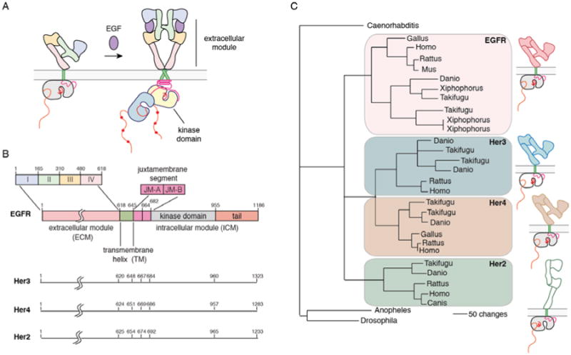
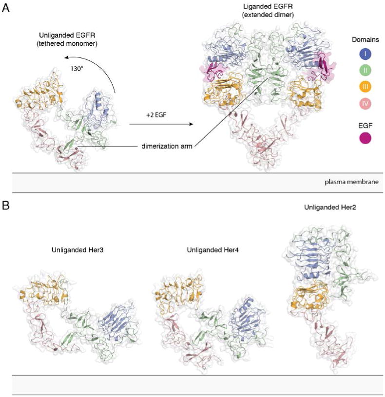
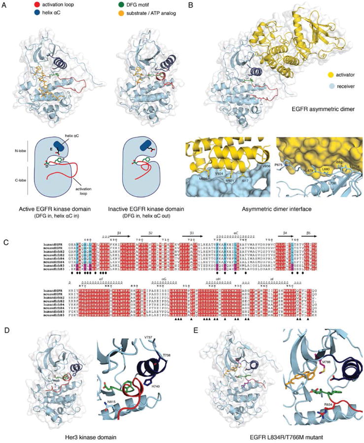
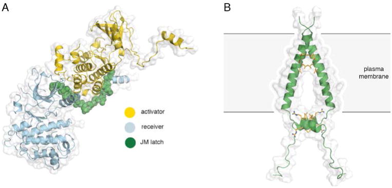
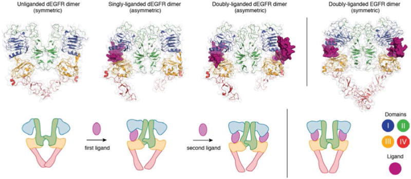
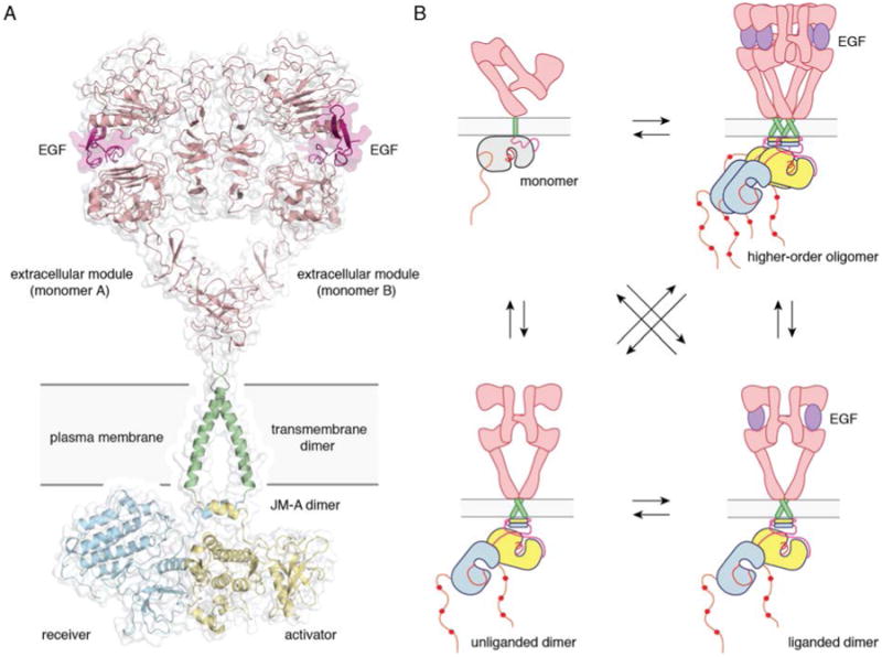
The epidermal growth factor receptor (EGFR) is a receptor tyrosine kinase that plays a critical role in the pathogenesis of many cancers. The structure of intact forms of this receptor has yet to be determined, but intense investigations of fragments of the receptor have provided a detailed view of its activation mechanism, which we review here. Ligand binding converts the receptor to a dimeric form, in which contacts are restricted to the receptor itself, allowing heterodimerization of the four EGFR family members without direct ligand involvement. Activation of the receptor depends on the formation of an asymmetric dimer of kinase domains, in which one kinase domain allosterically activates the other. Coupling between the extracellular and intracellular domains may involve a switch between alternative crossings of the transmembrane helices, which form dimeric structures. We also discuss how receptor regulation is compromised by oncogenic mutations and the structural basis for negative cooperativity in ligand binding.

Keywords: asymmetric dimer; ligand-induced dimerization; oncogenic mutations; receptor tyrosine kinase; transmembrane coupling.


Figures

Figure 1
Figure 1. Model for activation, domain architecture and evolutionary lineage of EGFR family members
A) Model for activation of EGFR family members. B) Domain boundaries in EGFR family members. C) Dendrogram of the evolution of EGFR family members (modified from (140)). Her2 does not have a known ligand (denoted by an open box), and the kinase domain of Her3 is defunctional (denoted by an open box).
Figure 2
Figure 2. Extracellular module structures for the EGFR family members
A) The conformational change induced by ligand binding. The tethered conformation of EGFR (left, PDB ID 1NQL, EGF bound at low pH was removed for clarity) rearranges to the extended conformation of EGFR (right, PDB ID 3NJP) upon ligand binding. B) Unliganded Her3 (PDB ID 1M6B) and Her4 (PDB ID 2AHX) can adopt a tethered conformation similar to EGFR, while Her2 (PDB ID 1N8H) is in an extended conformation, even in the absence of ligand.
Figure 3
Figure 3. Therapeutic antibodies target EGFR and Her2 in versatile ways
Structure of the EGFR-Cetuximab (PDB ID 1YY9), Her2-Pertuzumab (PDB ID 1S78), and Her2-Trastuzumab (PDB ID 1N8Z) complexes.
Figure 4
Figure 4. Kinase domain structures of the EGFR family members
A) The active (PDB ID 2GS6) and the inactive (PDB ID 2GS7) conformation of the EGFR kinase domain. Helix αC is colored blue, the DFG motif green, and the activation loop red. The active structure has an ATP analog - peptide conjugate bound, and the inactive structure has AMP-PNP bound (colored yellow). B) The asymmetric dimer of the EGFR kinase domain (PDB ID 2GS6). The activator kinase is colored yellow, and the receiver (enzymatically active) kinase is colored blue. Residue contacts important on the activator and the receiver are highlighted. C) A sequence alignment of the EGFR family members from human and mouse. Two regions containing the residues involved in the N- and C-lobe faces of the dimer interface are shown in the upper and lower panels, respectively. Identical residues are colored in red. Residues in the N- and C-lobe faces of the dimer interface are denoted by ovals and triangles, respectively. Blue and magenta highlight residues in the dimer interface that are conserved among EGFR, Her2, and Her4 but not in Her3. D) Structure of the Her3 kinase domain (PDB ID 3KEX) with a zoom-in of the active site, and residues resulting in a catalytically impaired kinase are highlighted. The catalytic Asp 813 in EGFR is replaced by Asn 815 in Her3, the critical Glu 738 in helix αC of EGFR is replaced with His 740 in Her3, and Val 737 and Thr 738 in Her3 stabilize the inactive conformation of helix αC. E) A structure of the L834R/T766M double mutant EGFR kinase domain (PDB ID 4LL0) with a zoom-in of the active site. The bound inhibitor, PD168393, is colored yellow, and the mutations are highlighted.
Figure 5
Figure 5. Structures of the juxtamembrane latch and the transmembrane helices of EGFR
A) The asymmetric dimer of EGFR with the juxtamembrane latch highlighted in green (PDB ID 3GOP). B) An NMR structure of the transmembrane and JM-A helices of EGFR in lipid bicelles (PDB ID 2M20). Interactions between the N-terminal GxxxG-like motifs on the transmembrane segments and the LRRLL motifs of the juxtamembrane segments are highlighted in yellow.
Figure 6
Figure 6. Structural basis for negative cooperativity in ligand binding to Drosophila. EGFR
The unliganded (PDB ID 3I2T), singly-liganded (PDB ID 3LTG) and doubly-liganded (PDB ID 3LTF) dEGFR dimer. The structure of the doubly liganded human EGFR dimer (PDB ID 3NJP) is shown on the right.
Figure 7
Figure 7. Full-length EGFR and its oligomerization states
A) A proposed composite model of full-length EGFR based on the structures of individual modules (PDB ID 3NJP for the extracellular module, PDB ID 2M20 for the transmembrane - JM-A helices, and PDB ID 2GS6 for the kinase domains). B) Schematics for possible oligomerization states of EGFR in cells.

References

    1. Ruiz-Trillo I, Burger G, Holland PW, King N, Lang BF, et al. The origins of multicellularity: a multi-taxon genome initiative. Trends in genetics : TIG. 2007;23:113–8. - PubMed
    1. Lim WA, Pawson T. Phosphotyrosine signaling: evolving a new cellular communication system. Cell. 2010;142:661–7. - PMC - PubMed
    1. Hubbard SR, Till JH. Protein tyrosine kinase structure and function. Annual review of biochemistry. 2000;69:373–98. - PubMed
    1. Avraham R, Yarden Y. Feedback regulation of EGFR signalling: decision making by early and delayed loops. Nat Rev Mol Cell Biol. 2011;12:104–17. - PubMed
    1. Endres NF, Engel K, Das R, Kovacs E, Kuriyan J. Regulation of the catalytic activity of the EGF receptor. Current opinion in structural biology. 2011;21:777–84. - PMC - PubMed

Publication types