Review
. 2015:10:371-93.
doi: 10.1146/annurev-pathol-012513-104644.

Diseases of pulmonary surfactant homeostasis

Affiliations
Review

Diseases of pulmonary surfactant homeostasis

Jeffrey A Whitsett et al. Annu Rev Pathol. 2015.

Abstract

Advances in physiology and biochemistry have provided fundamental insights into the role of pulmonary surfactant in the pathogenesis and treatment of preterm infants with respiratory distress syndrome. Identification of the surfactant proteins, lipid transporters, and transcriptional networks regulating their expression has provided the tools and insights needed to discern the molecular and cellular processes regulating the production and function of pulmonary surfactant prior to and after birth. Mutations in genes regulating surfactant homeostasis have been associated with severe lung disease in neonates and older infants. Biophysical and transgenic mouse models have provided insight into the mechanisms underlying surfactant protein and alveolar homeostasis. These studies have provided the framework for understanding the structure and function of pulmonary surfactant, which has informed understanding of the pathogenesis of diverse pulmonary disorders previously considered idiopathic. This review considers the pulmonary surfactant system and the genetic causes of acute and chronic lung disease caused by disruption of alveolar homeostasis.

Keywords: alveolar capillary dysplasia; alveolar proteinosis; interstitial lung disease; pulmonary alveolar microlithiasis; pulmonary fibrosis; respiratory distress syndrome.


Figures
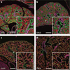
Figure 1
Figure 1
Stages of lung morphogenesis. Confocal microscopy was used to image lung architecture in the mouse from (a) day E14 (pseudoglandular) to (b) E16.5 (canalicular), (c) E18.5 (saccular), and (d) PN14 (alveolar period). TTF-1 (green) staining identifies epithelial cells; α-SMA (purple) identifies smooth muscle cells. Endomucin (red) marks pulmonary endothelial cells. Arrows indicate the regions of enlargement illustrated in panel insets. Scale bar = 200 µm for all panels. Immunofluorescent images courtesy of Dr. John Shannon, Department of Pediatrics, Cincinnati Children’s Hospital Medical Center and the University of Cincinnati, Cincinnati, Ohio. Abbreviations: α-SMA, α smooth muscle actin; E, embryonic; PN, postnatal; TTF-1, thyroid transcription factor-1.
Figure 2
Figure 2
Blueprint for specification and differentiation of the respiratory epithelium in the mouse, from day E3.5 to adult. The lungs are derived from progenitor cells located in the ventral region of the embryonic foregut. Endoderm is specified from neuroectoderm and mesoderm prior to the implantation of the embryo. This process is mediated in part by the expression of SOX17 and FOXA2, transcription factors critical for the formation of endoderm. Under the influence of WNT, SHH, FGF, and BMP signaling, the lung buds are distinguished from the gut tube and marked by the expression of NKX2-1 (thyroid transcription factor-1). The esophagus separates from the trachea, the latter marked by expression of SOX2, which is required for the formation and differentiation of airways and airway epithelial cells. Peripheral lung progenitors, expressing both NKX2-1 and SOX9, produce alveolar type I and type II epithelial cells. Surfactant proteins and lipids are produced after the differentiation of type II epithelial cells, which in preparation for birth are also under the control of NKX2-1, FOXA2, and associated transcription factors. Mutations in the genes encoding the transcription factors SOX2 and NKX2-1 cause pulmonary malformations; NKX2-1 regulates genes that are critical for alveolar homeostasis after birth. Abbreviations: Br, bronchiole; E, embryonic.
Figure 3
Figure 3
Biosynthesis of surfactant likely involves distinct pathways for surfactant proteins and lipids. SP-B and SP-C are trafficked from the endoplasmic reticulum to lamellar bodies via the Golgi complex and MVB; in contrast, surfactant phospholipids are likely directly transported from the endoplasmic reticulum to specific lipid importers (ABCA3) in the lamellar body–limiting membrane. Surfactant proteins and lipids are assembled into bilayer membranes that are secreted into the alveolar airspace, where they form a surface film at the air–liquid interface. Cyclical expansion and compression of the bioactive film results in the incorporation (large green arrow) and loss (red arrows) of lipids and proteins from the multilayered surface film. Surfactant components removed from the film are degraded in alveolar macrophages or are taken up by type II epithelial cells for recycling or degradation in the lysosome (red arrows). The MVB plays a key part in the integration of pathways for surfactant synthesis, recycling, and degradation. Abbreviations: ABCA3, ATP-binding cassette transporter A3; GM-CSF, granulocyte macrophage colony–stimulating factor; MVB, multivesicular body; PC, phosphatidylcholine; PG, phosphatidylglycerol; SP/SFTP, surfactant protein.
Figure 4
Figure 4
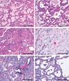
Histopathology of genetic disorders of surfactant homeostasis. (a) Histopathological specimen from the lung of a neonate with a lethal SFTPB mutation, demonstrating the typical pattern of pulmonary alveolar proteinosis composed of intraalveolar, foamy, eosinophilic, lipoproteinaceous material and thickened alveolar septa. (b) Histopathological specimen from the lung of an infant with a SFTPC mutation and chronic pneumonitis of infancy, showing muscularization of the thickened alveolar septa; patchy, intraalveolar, granular, proteinosis material; and alveolar type II cell hyperplasia. (c) Histopathological specimen from the lung of a neonate with a lethal ATP-binding cassette transporter A3 (ABCA3) mutation, demonstrating granular, eosinophilic, alveolar proteinosis material mixed with macrophages; thickened alveolar septa; and alveolar type II cell hyperplasia. (d) Histopathological specimen from the lung of a child with a mutation in the αchain of the granulocyte macrophage colony–stimulating factor (GM-CSF) receptor (CSF2RA), demonstrating both foamy and globular intraalveolar pulmonary alveolar proteinosis but with alveolar septa that appear thin and more normal. (e) Histopathological specimen from an infant with an NKX2-1 mutation, demonstrating features consistent with the genetic surfactant disorders including thickened, muscularized alveolar septa; intraalveolar accumulation of foamy macrophages (as seen in desquamative interstitial pneumonia); and hyperplastic alveolar type II cells. (f) Histopathological specimen from an infant with an NKX2-1 (TTF-1) mutation, demonstrating a mixed phenotype consisting of a diffuse alveolar growth abnormality that is superimposed on chronic interstitial inflammation and fibrosis and that resembles nonspecific interstitial pneumonia. All specimens are stained with hematoxylin and eosin; scale bars = 200 µm for all panels.
Figure 5
Figure 5
Histopathology of alveolar capillary dysplasia with misalignment of the pulmonary veins. Histopathological specimen from the lung of a neonate with a lethal FOXF1 mutation. (a) The pulmonary veins are large, dilated, congested vessels that are mislocated and lie adjacent to the pulmonary artery in the bronchoarterial compartment. (b) The pulmonary medial wall of the arteries is hypertrophic with increased immunostaining for α smooth muscle actin. (c) The pulmonary lobes and segments are hypoplastic, with abnormal formation of the parenchyma. There are decreased numbers of acinar and alveolar structures, with thickened interacinar and alveolar septa. The capillary bed is abnormal and has a decreased number of capillaries; these are disorganized and located primarily in the center of the thickened interstitial compartment (arrows). (d) Immunostaining for von Willebrand factor, a marker of endothelial cells, highlights the abnormal capillary network (arrows). Specimens in panels a and c were stained with hematoxylin and eosin; panels b and d were stained with immunoperoxidase and nuclear fast red counterstain. Scale bars for panels a and b = 200 µm; scale bars for panels c and d = 100 µm. Abbreviations: Art, artery; Br, bronchiole.

Similar articles

Cited by

References

    1. Metzger RJ, Klein OD, Martin GR, Krasnow MA. The branching programme of mouse lung development. Nature. 2008;453:745–750. - PMC - PubMed
    1. Perl AK, Wert SE, Nagy A, Lobe CG, Whitsett JA. Early restriction of peripheral and proximal cell lineages during formation of the lung. PNAS. 2002;99:10482–10487. - PMC - PubMed
    1. Morrisey EE, Hogan BL. Preparing for the first breath: genetic and cellular mechanisms in lung development. Dev. Cell. 2010;18:8–23. - PMC - PubMed
    1. Warburton D, El-Hashash A, Carraro G, Tiozzo C, Sala F, et al. Lung organogenesis. Curr. Top. Dev. Biol. 2010;90:73–158. - PMC - PubMed
    1. Kimura S, Hara Y, Pineau T, Fernandez-Salguero P, Fox CH, et al. The T/ebp null mouse: thyroid-specific enhancer-binding protein is essential for the organogenesis of the thyroid, lung, ventral forebrain, and pituitary. Genes Dev. 1996;10:60–69. - PubMed

Publication types

MeSH terms

Substances