

doi: 10.1371/journal.pbio.1002045.
eCollection 2015 Jan.
Adult neurogenesis in humans- common and unique traits in mammals
Affiliations
- PMID: 25621867
- PMCID: PMC4306487
- DOI: 10.1371/journal.pbio.1002045
Item in Clipboard
Adult neurogenesis in humans- common and unique traits in mammals
PLoS Biol.
.
Abstract
New neurons are continuously generated in specific regions in the adult brain. Studies in rodents have demonstrated that adult-born neurons have specific functional features and mediate neural plasticity. Data on the extent and dynamics of adult neurogenesis in adult humans are starting to emerge, and there are clear similarities and differences compared to other mammals. Why do these differences arise? And what do they mean?
Conflict of interest statement
The authors have declared that no competing interests exist.
Figures
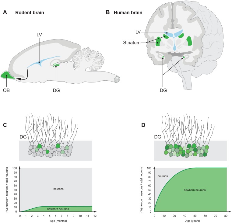
New neurons are indicated in green. (A) Neuroblasts that are generated in the subventricular zone lining the lateral ventricle (LV) in rodents migrate to the OB, a structure crucial for olfaction, where they integrate as interneurons. (B) Neuroblasts are present in the subventricular zone also in humans, and new neurons integrate in the adjacent striatum, which plays an essential role in movement coordination, procedural learning, and memory, as well as motivational and emotional control. New neurons are continuously generated in the DG of the hippocampus—a brain structure essential for memory and mood control—in both rodents and humans (A, B). A limited subpopulation of DG neurons are subject to exchange in rodents (C), whereas the majority turn over in humans (D) [–6]. The neurons within the turning over population are continuously exchanged. A value of 100% on the y-axis means that all neurons have been replaced since the individual’s birth.
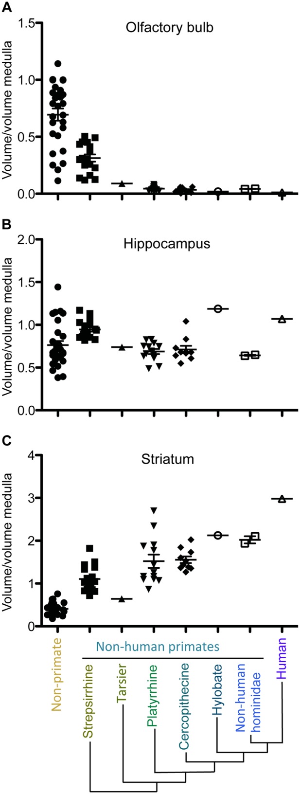
Species are grouped according to major phylogenetic classes varying in phylogenetic distance from humans: nonprimates first (e.g., shrews, tenrecs, hedgehogs), then Strepsirrhine (e.g., lemur), Tarsier, Platyirrhine (e.g., New World monkey), Cercopithecine (e.g., baboon, macaque), Hylobate (e.g., gibbon), Nonhuman hominidae (e.g., chimpanzee, gorilla). The proportional volume of the OBs decreases across primate species. Humans display the most pronounced reduction in OB volume. Hippocampal volumes appear to maintain their proportions across species, whereas proportional striatal volumes increase with evolution. The proportions of regional brain volumes are calculated as proportions of medulla volumes, because no grade shifts in the relationship between medulla volume and body size are observed [34]. Volumetric measurements are from Stephan et al. [48].
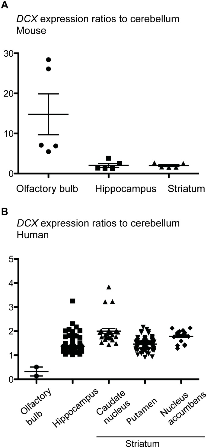
In mice, DCX expression is much higher in the OB than in the hippocampus and striatum. In humans, only background levels are detected in the OB, whereas higher DCX expression levels are reached in the human hippocampus and striatum. mRNA expression was measured by in situ hybridization, expression profiling, and RNA sequencing. Data are from geo (GSE 2361, GSE 45878, GSE 46706, GSE 1133, GDS1490, GDS182) and from the Allen Brain Atlas. The data points for the human OB show pooled values for several donors.
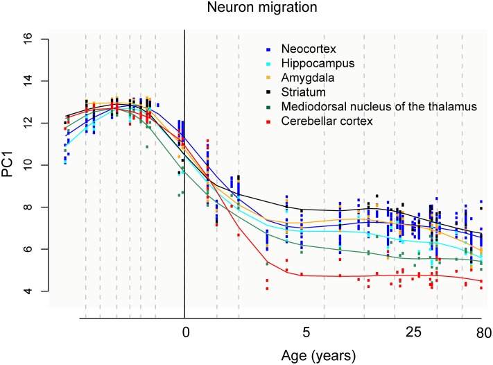
Y-axis, first principal component (PC) value for gene expression. Expression levels of 100 genes reported to be associated with neuronal migration are taken into account (see Kang et al. for details on the statistical methods for the principal component analysis and exhaustive list of genes included). X-axis, subject age in years. Data from Kang et al. [35].
References
Publication types
MeSH terms
LinkOut - more resources
Full Text Sources
Other Literature Sources
