Review
. 2015 Feb:32:82-91.
doi: 10.1016/j.ceb.2015.01.001. Epub 2015 Jan 23.

Intermediate filament mechanics in vitro and in the cell: from coiled coils to filaments, fibers and networks

Affiliations
Review

Intermediate filament mechanics in vitro and in the cell: from coiled coils to filaments, fibers and networks

Sarah Köster et al. Curr Opin Cell Biol. 2015 Feb.

Abstract

Intermediate filament proteins form filaments, fibers and networks both in the cytoplasm and the nucleus of metazoan cells. Their general structural building plan accommodates highly varying amino acid sequences to yield extended dimeric α-helical coiled coils of highly conserved design. These 'rod' particles are the basic building blocks of intrinsically flexible, filamentous structures that are able to resist high mechanical stresses, that is, bending and stretching to a considerable degree, both in vitro and in the cell. Biophysical and computer modeling studies are beginning to unfold detailed structural and mechanical insights into these major supramolecular assemblies of cell architecture, not only in the 'test tube' but also in the cellular and tissue context.


Figures

Figure 1
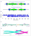
Figure 1. IF protein organization
A. Domain organization of lamin A and vimentin as representatives for nuclear and cytoplasmic IF proteins: A central α-helical “rod” is flanked by non-α-helical “head” and “tail” domains. Boxes represent amino acid sequence segments engaged in coiled-coil (green) or “paired bundle” (yellow) formation; the IF-consensus motifs are indicated in blue. The “linker” segments between coil 1A and coil 1B as well as those between coil 1B and coil 2 may be α-helical in the case of lamins, but are probably of unique fold in cytoplasmic IF proteins. The circle in the lamin tail represents an Ig fold. B. Charge distribution of vimentin demonstrating the basic nature of the rod and the rather dense pattern of alternating basic and acidic charges along the rod. C. Model of a vimentin coiled-coil dimer. Vimentin-like IF proteins exhibit pre-coil domains probably adopting an α-helical fold (PCD) not seen in lamins and keratins (redrawn from Ref. 6). Paired bundles are in orange, the “linker” domains are designated L1 and L12. The numbers in brackets indicate the number of amino acids in the “heads” and “tails”, respectively. D. Potential hetero-coiled-coil formation exhibited by two coiled-coil dimers in the ~3 nm head-to-tail overlap region directly observed for lamins (for details see Ref. 13).
Figure 2
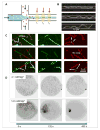
Figure 2. Vimentin in vitro assembly
A. Vimentin assembly in a diffusive mixing device. Assembly buffer injected from the two side channels are mixed diffusively with vimentin tetramers (green bars) injected from the main channel. As a result of the increased ionic strength, higher order complexes and eventually unit-length filaments form (green blocks at t2 and t3). The red arrows indicate examples for measurement positions, by e.g. x-ray scattering or fluorescence spectroscopy, which correspond to different time points in the assembly process (adapted from Ref. 7). B. Individual vimentin IF confined in microchannels. The width of the channels was 1.2 μm, 1.6 μm and 2.7 μm, top to bottom (adapted from Ref. 18). C. Subunit exchange along mature filaments. The overlay of the red and the green channel shows different modes of interaction between differently labeled subunits: 1 – end-to-end annealing; 2 – overlapping of filaments; 3 – exchanged subunits (adapted from Ref. 22). D. Impact of divalent ions on vimentin filament networks. Pre-assembled vimentin IFs were challenged with different concentrations of magnesium in microfluidic drops: above ~ 10 mM Mg2+ the networks aggregate strongly (adapted from Ref. 32).
Figure 3
Figure 3. In vivo IF mechanics
A. Data from optical stretcher experiments show that wild type keratinocytes are less deformable than keratin knockout cells (J(t) shows creep deformation; from Ref. 46). B. Atomic force measurements; shown are stiffness maps of a live wild type keratinocyte (left) and a keratin knock-out cell (right); scale bar 10 μm (from Ref. 47). C. Pressurization of lobopodia by the “nuclear piston”. (Inset) Migration of primary human fibroblasts in a 3D extracellular matrix. (Main figure) Nesprin 3 connects the nucleus via plectin to intermediate filaments and actomyosin contractility. These connections help pull the nucleus forward to pressurize the forward cytoplasmic compartment and sustain high-pressure lobopodia-based 3D motility; from Ref. . D. Buckling event in a network of keratin bundles in a SW13 cell stably transfected with CFP-K8 and YFP-K18. (Left) two time frames, 2 s apart, showing the buckling event. (Right) marked ROI (region of interest) as shown on the left hand side.

Similar articles

Cited by

References

    1. Parry DAD, Steinert M. Intermediate filaments: molecular architecture, assembly, dynamics and polymorphism. Quart Rev Biophys. 1999;32:99–187. - PubMed
    1. Erber A, Riemer D, Hofemeister H, Bovenschulte M, Stick R, Panopoulou G, Lehrach H, Weber K. Characterization of the Hydra lamin and its gene: A molecular phylogeny of metazoan lamins. J Mol Evol. 1999;49:260–271. - PubMed
    1. Herrmann H, Aebi U. Intermediate Filaments: Molecular Structure, Assembly Mechanism, and Integration Into Functionally Distinct Intracellular Scaffolds. Ann Rev Biochem. 2004;73:749–789. - PubMed
    1. Herrmann H, Kreplak L, Aebi U. Isolation, characterization, and in vitro assembly of intermediate filaments. Methods Cell Biol. 2004;78:3–24. - PubMed
    1. Herrmann H, Häner M, Brettel M, Müller SA, Goldie KN, Fedtke B, Lustig A, Franke WW, Aebi U. Structure and Assembly Properties of the Intermediate Filament Protein Vimentin: The Role of its Head, Rod and Tail Domains. J Mol Biol. 1996;264:933–953. - PubMed

Publication types