. 2015 Apr;197(7):1197-207.
doi: 10.1128/JB.02508-14. Epub 2015 Jan 26.

Structure-function analysis of VapB4 antitoxin identifies critical features of a minimal VapC4 toxin-binding module

Affiliations

Structure-function analysis of VapB4 antitoxin identifies critical features of a minimal VapC4 toxin-binding module

Guangze Jin et al. J Bacteriol. 2015 Apr.

Abstract

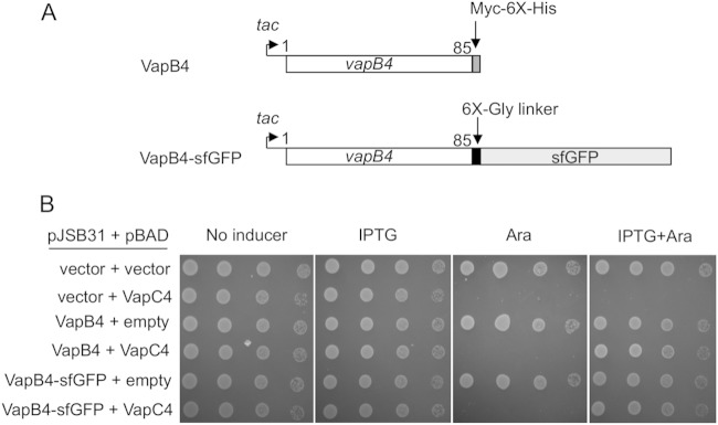
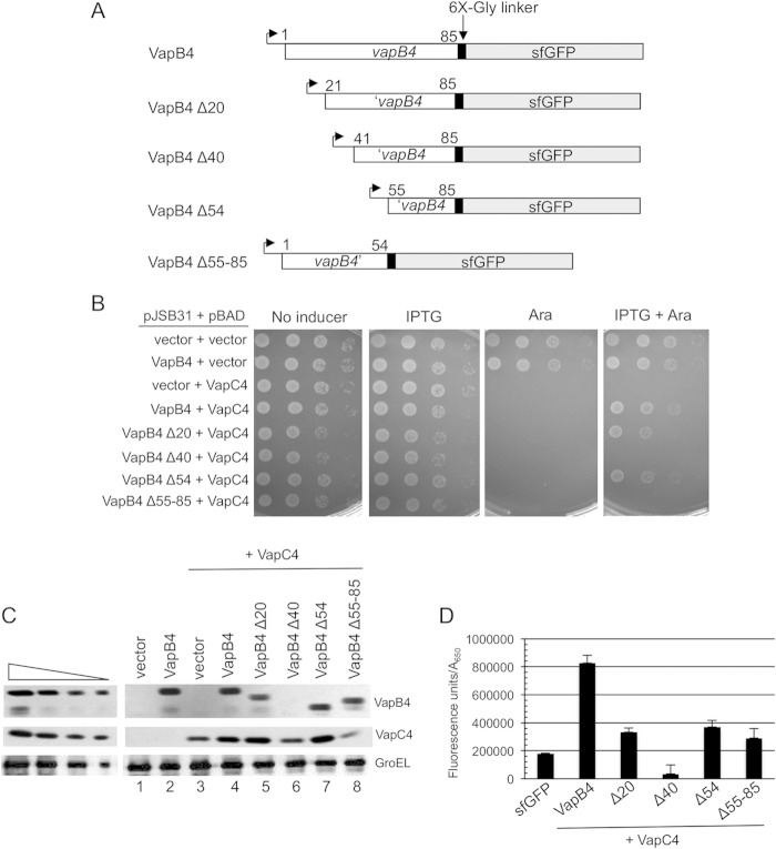
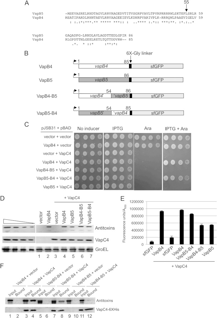
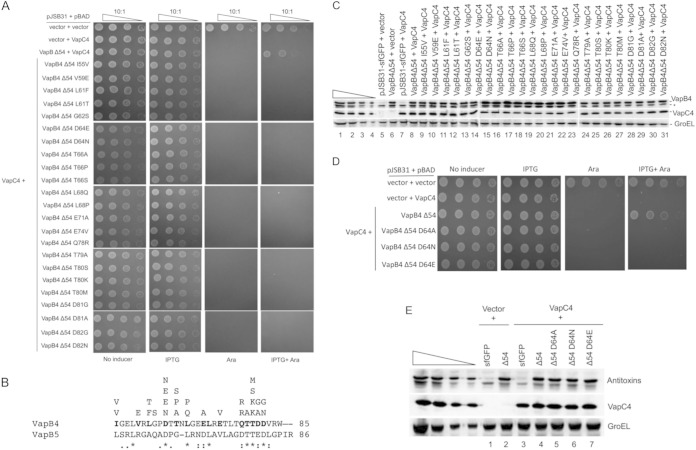
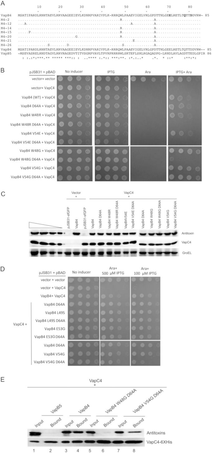
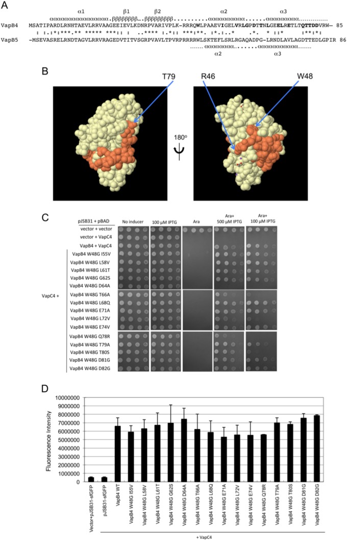
Bacterial toxin-antitoxin systems play a critical role in the regulation of gene expression, leading to developmental changes, reversible dormancy, and cell death. Type II toxin-antitoxin pairs, composed of protein toxins and antitoxins, exist in nearly all bacteria and are classified into six groups on the basis of the structure of the toxins. The VapBC group comprises the most common type II system and, like other toxin-antitoxin systems, functions to elicit dormancy by inhibiting protein synthesis. Activation of toxin function requires protease degradation of the VapB antitoxin, which frees the VapC toxin from the VapBC complex, allowing it to hydrolyze the RNAs required for translation. Generally, type II antitoxins bind with high specificity to their cognate toxins via a toxin-binding domain and endow the complex with DNA-binding specificity via a DNA-binding domain. Despite the ubiquity of VapBC systems and their critical role in the regulation of gene expression, few functional studies have addressed the details of VapB-VapC interactions. Here we report on the results of experiments designed to identify molecular determinants of the specificity of the Mycobacterium tuberculosis VapB4 antitoxin for its cognate VapC4 toxin. The results identify the minimal domain of VapB4 required for this interaction as well as the amino acid side chains required for binding to VapC4. These findings have important implications for the evolution of VapBC toxin-antitoxin systems and their potential as targets of small-molecule protein-protein interaction inhibitors.

Importance: VapBC toxin-antitoxin pairs are the most widespread type II toxin-antitoxin systems in bacteria, where they are thought to play key roles in stress-induced dormancy and the formation of persisters. The VapB antitoxins are critical to these processes because they inhibit the activity of the toxins and provide the DNA-binding specificity that controls the synthesis of both proteins. Despite the importance of VapB antitoxins and the existence of several VapBC crystal structures, little is known about their functional features in vivo. Here we report the findings of the first comprehensive structure-function analysis of a VapB toxin. The results identify the minimal toxin-binding domain, its modular antitoxin function, and the specific amino acid side chains required for its activity.


Figures

FIG 1
FIG 1
Expression of the VapB4 (Rv0596c)-myc-6× His or VapB4-sfGFP fusion in trans inhibits the toxicity of VapC4 (Rv0595c). (A) The open reading frame of VapB4 from M. tuberculosis was cloned into the expression plasmid pJSB31 or pJSB31-sfGFP, creating a myc-6× His fusion or a sfGFP fusion, respectively. (B) The growth of E. coli strains carrying the indicated plasmids was compared using the serial dilution cell spotting assay, as described in Materials and Methods.
FIG 2
FIG 2
The C-terminal region of VapB4 (positions 55 to 85) is sufficient for VapB4 antitoxin activity. (A) The open reading frames of VapB4 and its deletion mutants were cloned into the pJSB31-sfGFP plasmid, creating sfGFP fusions. (B) Spotting assay for VapB4 deletion mutants carried out as described in the legend to Fig. 1B. (C) Western blot analysis of cells carrying pJSB31-sfGFP (vector) or pJSB31 carrying the indicated VapB4 alleles and pBAD-VapC4. The cells were grown in M9-glycerol (0.2%) medium at 37°C and induced with 0.02% l-arabinose and 500 μM IPTG for 30 min. Blots were probed with anti-GFP antibody for VapB4 or its mutant proteins and anti-myc antibody for VapC4-myc proteins. Anti-GroEL served as a loading control. The four lanes in each of the panels on the left show a 2-fold dilution series of VapB4-sfGFP, which served as a control for signal linearity. (D) E. coli strain LMG194 carrying the indicated plasmids was grown in M9-glycerol (0.2%) medium supplemented with 50 μg/ml ampicillin and 30 μg/ml chloramphenicol at 37°C to an A600 of 0.1 to 0.2 and induced with 0.02% l-arabinose and 500 μM IPTG for 2 h. GFP fluorescence was measured in a Typhoon 9410 imager and normalized to the OD600 of the culture. Error bars illustrate the standard deviations of triplicate measurements.
FIG 3
FIG 3
VapB5-VapB4 but not VapB4-VapB5 functions as an antitoxin to VapC4. (A) ClustalW alignment of VapB4 (Rv0596c) with the VapB5 (Rv0626) sequence. Colons, nonidentical residues; periods, functionally similar residues; asterisks, identical residues. (B) The open reading frames of VapB4, VapB5, and their domain-swapped mutants were cloned into the expression plasmid pJSB31-sfGFP, creating sfGFP fusions. (C) Spotting assay for E. coli strain LMG194 carrying the indicated plasmids carried out as described in the legend to Fig. 1B. (D) Western blot analysis was performed as described in the legend to Fig. 2C. (E) Fluorescence measurements for VapB4, VapB5, and their domain-swapped mutants were performed as described in the legend to Fig. 2D. (F) Western blot analysis of copurified proteins from E. coli LMG194 cells carrying plasmids expressing the indicated VapB combinations and VapC4-myc-6× His. Cells were induced with 0.02% l-arabinose and 500 μM IPTG for 30 min at 37°C. Western blots were probed with anti-GFP antibody for VapB4 or its mutant proteins and anti-myc antibody for VapC4 proteins.
FIG 4
FIG 4
Single mutations disrupt the antitoxin activity of VapB4 Δ54. (A) Spotting assay for E. coli strain LMG194 carrying the indicated plasmids carried out as described in the legend to Fig. 1B. (B) Single amino acid changes in the C-terminal 31-amino-acid domain of VapC4 and comparison with the same portion of VapB5. Each amino acid residue listed above VapB4 protein sequence represents a single mutation isolated from the screen. (C) Western blot analysis was carried out as described in the legend to Fig. 2C. The asterisk marks the position of an anti-GFP-positive band. (D) Spotting assay for E. coli strain LMG194 carrying the indicated plasmids carried out as described in the legend to Fig. 1B. (E) Western blot analysis was carried out as described in the legend to Fig. 2C.
FIG 5
FIG 5
Mutation of W48 results in the loss of antitoxin activity of full-length VapC4 D64A. (A) Comparison of the sequences of VapB4 with those of the defective mutants isolated from screening. (B) Spotting assay for E. coli strain LMG194 carrying the indicated plasmids carried out as described in the legend to Fig. 1B. (C) Western blot analysis was carried out as described in the legend to Fig. 2C. (D) E. coli strain LMG194 carrying the indicated plasmids was grown and spotted with 10-fold dilutions on M9 medium supplemented with 100 μg/ml ampicillin, 30 μg/ml chloramphenicol, and 0.2% glycerol in the presence or absence of 0.02% l-arabinose and 500 μM (or 100 μM) IPTG and grown for 16 h at 37°C. (E) Western blot analysis of copurified proteins from E. coli LMG194 cells carrying plasmids expressing VapB5 or the indicated VapB4 alleles and VapC4-myc-6× His. Cells were induced with 0.02% l-arabinose and 500 μM IPTG for 30 min at 37°C. Western blots were probed with anti-GFP antibody for VapB4 or its mutant proteins and anti-myc antibody for VapC4 proteins.
FIG 6
FIG 6
Mutation of W48 results in the loss of antitoxin activity of full-length VapC4 D64A. (A) Amino acid sequence alignment of VapB4 and VapB5, with their corresponding predicted secondary structure elements assigned. Secondary structure assignments for VapB4 were from the Phyre2 server (43), and those for VapB5 were from reference . (B) Structure of the VapB5 (orange)-VapC5 (white) heterodimer (37). The structure with PDB accession number 3DBO was generated by use of the PISA program (44). (C) Spotting assay for E. coli strain LMG194 carrying the indicated plasmids carried out as described in the legend to Fig. 5D. (D) Fluorescence measurements for VapB4 mutants that were assayed for growth and for which the results are presented in panel C. Measurements were taken directly from the third dilution spot on the plate with 100 μM IPTG after determination that all spots in the dilution were in the linear range of detection of the Typhoon 9410 imager. The columns report the average fluorescence intensity from three biological replicates, and error bars illustrate standard deviations. WT, wild type.

References

    1. Gerdes K, Molin S. 1986. Partitioning of plasmid R1. Structural and functional analysis of the parA locus. J Mol Biol 190:269–279. - PubMed
    1. Ogura T, Hiraga S. 1983. Mini-F plasmid genes that couple host cell division to plasmid proliferation. Proc Natl Acad Sci U S A 80:4784–4788. doi:10.1073/pnas.80.15.4784. - DOI - PMC - PubMed
    1. Gerdes K. 2000. Toxin-antitoxin modules may regulate synthesis of macromolecules during nutritional stress. J Bacteriol 182:561–572. doi:10.1128/JB.182.3.561-572.2000. - DOI - PMC - PubMed
    1. Hayes F. 2003. Toxins-antitoxins: plasmid maintenance, programmed cell death, and cell cycle arrest. Science 301:1496–1499. doi:10.1126/science.1088157. - DOI - PubMed
    1. Aakre CD, Phung TN, Huang D, Laub MT. 2013. A bacterial toxin inhibits DNA replication elongation through a direct interaction with the beta sliding clamp. Mol Cell 52:617–628. doi:10.1016/j.molcel.2013.10.014. - DOI - PMC - PubMed

Publication types

MeSH terms

Associated data

LinkOut - more resources