. 2015 Jan;17(1):32-42.
doi: 10.1016/j.neo.2014.11.001.

Recombinant interleukin-1 receptor antagonist conjugated to superparamagnetic iron oxide nanoparticles for theranostic targeting of experimental glioblastoma

Affiliations

Recombinant interleukin-1 receptor antagonist conjugated to superparamagnetic iron oxide nanoparticles for theranostic targeting of experimental glioblastoma

Maxim A Shevtsov et al. Neoplasia. 2015 Jan.

Abstract

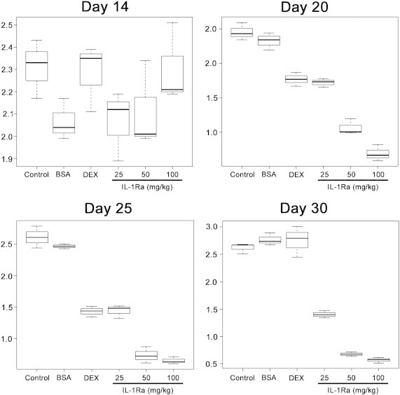
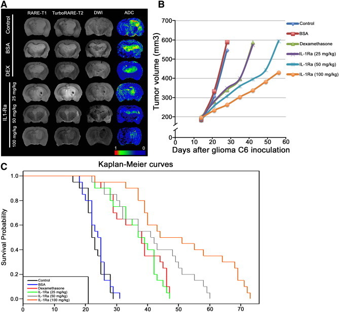
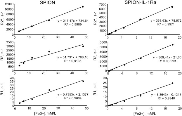
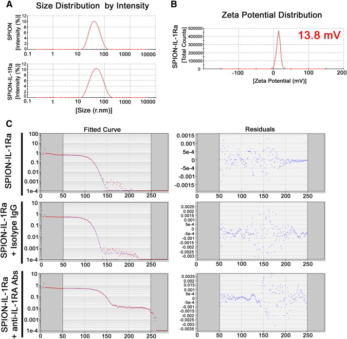
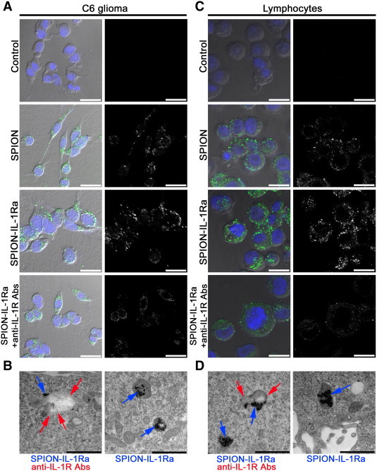
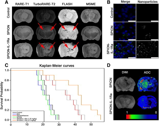
Cerebral edema commonly accompanies brain tumors and contributes to neurologic symptoms. The role of the interleukin-1 receptor antagonist conjugated to superparamagnetic iron oxide nanoparticles (SPION-IL-1Ra) was assessed to analyze its anti-edemal effect and its possible application as a negative contrast enhancing agent for magnetic resonance imaging (MRI). Rats with intracranial C6 glioma were intravenously administered at various concentrations of IL-1Ra or SPION-IL-1Ra. Brain peritumoral edema following treatment with receptor antagonist was assessed with high-field MRI. IL-1Ra administered at later stages of tumor progression significantly reduced peritumoral edema (as measured by MRI) and prolonged two-fold the life span of comorbid animals in a dose-dependent manner in comparison to control and corticosteroid-treated animals (P < .001). Synthesized SPION-IL-1Ra conjugates had the properties of negative contrast agent with high coefficients of relaxation efficiency. In vitro studies of SPION-IL-1Ra nanoparticles demonstrated high intracellular incorporation and absence of toxic influence on C6 cells and lymphocyte viability and proliferation. Retention of the nanoparticles in the tumor resulted in enhanced hypotensive T2-weighted images of glioma, proving the application of the conjugates as negative magnetic resonance contrast agents. Moreover, nanoparticles reduced the peritumoral edema confirming the therapeutic potency of synthesized conjugates. SPION-IL-1Ra nanoparticles have an anti-edemal effect when administered through a clinically relevant route in animals with glioma. The SPION-IL-1Ra could be a candidate for theranostic approach in neuro-oncology both for diagnosis of brain tumors and management of peritumoral edema.


Figures

Figure 1
Figure 1
Confocal microscopy of the C6 glioma following i.v. administration of the IL-1Ra. Nuclei were stained with DAPI (blue). IL-1Ra was detected with monoclonal antibodies conjugated with Alexa Fluor 555 (red). Scale bar, 75 μm.
Figure 2
Figure 2
Side-by-side boxplots of ADC. ADC was calculated for the five experimental groups and for the control group on days 14, 20, 25, and 30 following C6 glioma cell inoculation.
Figure 3
Figure 3
Anti-edemal activity of recombinant IL-1Ra and animal survival analysis. (A) MRI of the C6 glioma at day 25 for the animals from control and treated with BSA, dexamethasone (DEX), and IL-1Ra at 25, 50, and 100 mg/kg. MR scans were obtained at RARE-T1, TurboRARE-T2, and DWI regimens. Additionally for each animal, the ADC map was calculated. (B) Tumor volume growth dynamics (mm3) for animals from five experimental groups and control group. (C) Kaplan-Meier survival curves for control and experimental groups.
Figure 4
Figure 4
Magnetic relaxation rates R2*, R2, and R1 of water protons in SPIONs and SPION–IL-1Ra conjugate dispersion in dependence on the Fe concentration at 20°C.
Figure 5
Figure 5
Characterization of the synthesized SPION–IL-1Ra conjugates. (A) Hydrodynamic size (r.nm) of the SPIONs and SPION–IL-1Ra nanoparticles. (B) Zeta potential for the SPION–IL-1Ra conjugates. (C) DLS assay for the SPION–IL-1Ra particles incubated with isotype IgG antibodies or monoclonal anti–IL-1Ra antibodies for 4 hours.
Figure 6
Figure 6
Interaction of the magnetic conjugates of IL-1Ra with C6 cells or lymphocytes. (A) Confocal microscopy images of the C6 cells following 24 hours of incubation with PBS, SPIONs (150 μg/ml), and SPION–IL-1Ra conjugates (150 μg/ml). Nuclei were stained with DAPI (blue). Magnetic nanoparticles were detected by reflecting laser scanning at 488 nm (green). Scale bar, 25 μm. For analysis of the receptor-mediated endocytosis, C6 cells were also incubated with anti–IL-1R monoclonal antibodies. (B) TEM of the C6 cell incubated for 24 hours with SPION–IL-1Ra conjugates. Electron dense nanoparticles were present in the cytoplasm of cells in the endosome-like structures (blue solid arrow). Receptors for IL-1 were detected with monoclonal anti–IL-1R antibodies (red solid arrow). Scale bar, 500 nm. (C) Confocal microscopy images of the rat lymphocytes following 24 hours of incubation with PBS, SPIONs (150 μg/ml), and SPION–IL-1Ra conjugates (150 μg/ml). Nuclei were stained with DAPI (blue). Magnetic nanoparticles are represented in the cytoplasm as green dots. Scale bar, 25 μm. For analysis of the receptor-mediated endocytosis, C6 cells were also incubated with anti–IL-1R monoclonal antibodies. (D) TEM of the lymphocyte incubated for 24 hours with SPION–IL-1Ra conjugates (blue arrows). Receptors for IL-1 were detected with monoclonal anti–IL-1R antibodies (red arrows). Scale bar, 500 nm.
Figure 7
Figure 7
C6 glioma targeting with SPION–IL-1Ra conjugates. (A) MRI of the C6 glioma 24 hours following i.v. treatment with SPIONs or SPION–IL-1Ra conjugates. MR scans were obtained at RARE-T1, TurboRARE-T2, FLASH, and MSME regimens. Retention of the magnetic nanoparticles is presented as hypotensive zones at T2-weighted images and in the regimen of the gradient echo (red arrows). (B) Confocal microscopy of the C6 glioma sections. Nanoparticles were detected by reflecting laser scanning (green) and nuclei were stained with DAPI (blue). Scale bar, 75 μm. (C) Kaplan-Meier survival curves for the control group and animals treated with BSA, dexamethasone, and SPION–IL-1Ra conjugates at 1.25 and 2.5 mg/kg of IL-1Ra. (D) DWIs and the corresponding ADC maps for the animals treated with SPION or SPION–IL-1Ra conjugates.

Similar articles

Cited by

References

    1. Schoenegger K., Oberndorfer S., Wuschitz B., Struhal W., Hainfellner J., Prayer D., Heinzl H., Lahrmann H., Marosi C., Grisold W. Peritumoral edema on MRI at initial diagnosis: an independent prognostic factor for glioblastoma? Eur J Neurol. 2009;16:874–878. - PubMed
    1. Koehler P.J. Use of corticosteroids in neuro-oncology. Anticancer Drugs. 1995;16:19–33. - PubMed
    1. Dietrich J., Rao K., Pastorino S., Kesari S. Corticosteroids in brain cancer patients: benefits and pitfalls. Expert Rev Clin Pharmacol. 2011;4:233–242. - PMC - PubMed
    1. Roth P., Regli L., Tonder M., Weller M. Tumor-associated edema in brain cancer patients: pathogenesis and management. Expert Rev Anticancer Ther. 2013;13:1319–1325. - PubMed
    1. Badie B., Schartner J.M., Hagar A.R., Prabakaran S., Peebles T.R., Bartley B., Lapsiwala S., Resnick D.K., Vorpahl J. Microglia cyclooxygenase-2 activity in experimental gliomas: possible role in cerebral edema formation. Clin Cancer Res. 2003;9:872–877. - PubMed

Publication types

MeSH terms