. 2015 Jan;17(1):141-51.
doi: 10.1016/j.neo.2014.12.002.

RhoC mediates epidermal growth factor-stimulated migration and invasion in head and neck squamous cell carcinoma

Affiliations

RhoC mediates epidermal growth factor-stimulated migration and invasion in head and neck squamous cell carcinoma

Zohra Tumur et al. Neoplasia. 2015 Jan.

Abstract

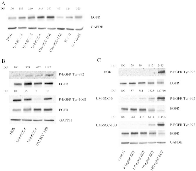
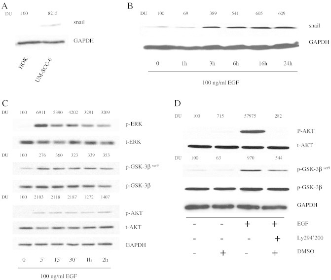
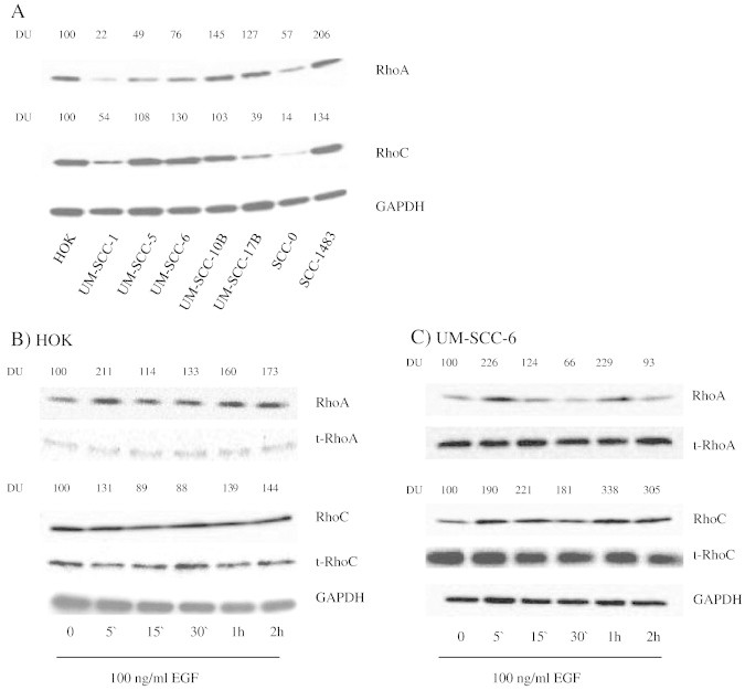
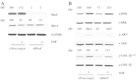
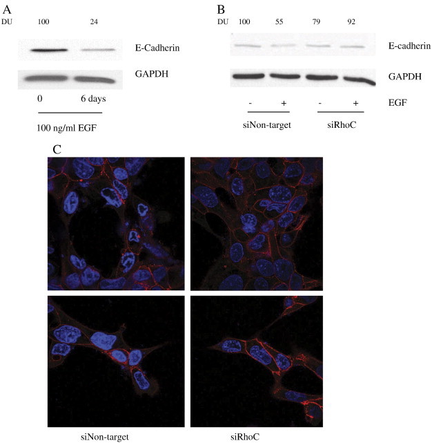
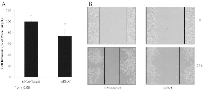
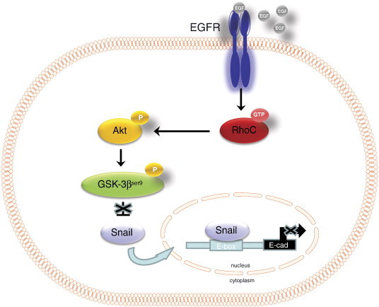
Epidermal growth factor receptor (EGFR) is overexpressed in head and neck squamous cell carcinoma (HNSCC) where it has been shown to promote tumor cell invasion upon phosphorylation. One mechanism by which EGFR promotes tumor progression is by activating signal cascades that lead to loss of E-cadherin, a transmembrane glycoprotein of the cell-cell adherence junctions; however mediators of these signaling cascades are not fully understood. One such mediator, RhoC, is activated upon a number of external stimuli, such as epidermal growth factor (EGF), but its role as a mediator of EGF-stimulated migration and invasion has not been elucidated in HNSCC. In the present study, we investigate the role of RhoC as a mediator of EGF-stimulated migration and invasion in HNSCC. We show that upon EGF stimulation, EGFR and RhoC were strongly activated in HNSCC. This resulted in activation of the phosphatidylinositol 3-Kinase Akt pathway (PI3K-Akt), phosphorylation of GSK-3β at the Ser(9) residue, and subsequent down regulation of E-cadherin cell surface expression resulting in increased tumor cell invasion. Knockdown of RhoC restored E-cadherin expression and inhibited EGF-stimulated migration and invasion. This is the first report in HNSCC demonstrating the role RhoC plays in mediating EGF-stimulated migration and invasion by down-regulating the PI3K-Akt pathway and E-cadherin expression. RhoC may serve as a treatment target for HNSCC.


Figures

Figure 1
Figure 1
EGFR is expressed and phosphorylated upon EGF binding in HNSCC cell lines. (A) Whole cell lysate of HOK, UM-SCC-(1,5,6,10B, 17B), and SCC-(0, 1483) were separated by PAGE and blotted with EGFR antibodies. The same membrane was subsequently blotted with GAPDH antibody to verify equivalency of loading. DU = densitometic units. (B) HOK and UM-SCC-5, -6, and -10B cells were analyzed for endogenous expression of phospho-EGFR at Tyr992 and phospho-EGFR at Tyr1068. Total EGFR and GAPDH were used as loading controls. (C) Dose response of EGF-mediated phosphorylation of EGFR. HOK, UM-SCC-6, and -10B cells were stimulated with 1 to 100 ng/ml of EGF for 5 min and then were analyzed for EGFR-Tyr 992 phosphorylation.
Figure 2
Figure 2
EGFR phosphorylation at Tyr992 activates the PI3-kinase pathway and up-regulates Snail protein expression. (A) Snail expression in HOK and UM-SCC-6 was examined via immunoblot analysis. (B) Snail expression is up-regulated following EGF stimulation. UM-SCC-6 cells were stimulated with 100 ng/ml of EGF for varying durations from 1 to 24 hours. Whole cell lysates were blotted to detect Snail expression. GAPDH was used as loading control. (C) ERK, GSK-3βser9 and AKT are phosphorylated following EGF stimulation. UM-SCC-6 cells were stimulated with 100 ng/ml EGF for 5 min to 2 h. Whole cell lysates were blotted with phospho-ERK, phospho-GSK-3βser9 and phospho-AKT antibodies followed by total ERK, AKT and GSK-3βser9 antibodies, respectively. (D) EGFR phosphorylates GSK-3βser9 via the PI3-kinase pathway. UM-SCC-6 cells were pre- incubated for 1 hour with 25 μM of LY294002 or DMSO as vehicle control followed by 100ng/ml EGF stimulation. Akt and GSK-3βser9 activation were evaluated by immunoblot analysis of whole cell lysates with phospho-Akt and phosphor-GSK-3βser9, respectively. The membrane was subsequently blotted with total Akt, GSK-3βser9, and GAPDH antibodies as loading controls.
Figure 3
Figure 3
RhoC is expressed in HNSCC and is activated by EGFR. (A) Whole cell lysate of HOK, UM-SCC-(1, 5, 6, 10B, 17B), and SCC-(0, 1483) were separated by PAGE and blotted with RhoA and RhoC antibodies. GAPDH was used as loading control. (B) HOK and UM-SCC-6 cells were stimulated with 100 ng/ml of EGF for varying durations from 5 minutes to 2 hours and RhoA and RhoC activation was evaluated by a pull down assay. Total RhoA, RhoC and GAPDH were used as loading controls. Active, GTP-bound Rho was quantified by densitometry and normalized to total normalized Rho with respect to GAPDH.
Figure 4
Figure 4
RhoC knock-down decreases EGF-stimulated Akt and GSK-3βser9 phosphorylation. (A) Expression of RhoC in HNSCC cells transfected with siRhoC. Plasmid expressing either control siRNA (siNon-target) or RhoC-specific siRNA (siRhoC) was transfected into UM-SCC-6 cells as described in the Materials and Methods. The cells were then simulated with 100 ng/ml EGF. RhoA and RhoC expression were confirmed by immunoblot analysis with GAPDH serving as a loading control. (B) At 72 hours following transfection with either siNon-target or siRhoC, UM-SCC-6 cells were stimulated with 100 ng/ml EGF or vehicle control for 5 minutes. Whole cell lysates were then blotted with phospho-ERK, phospho-GSK-3βser9 and phospho-AKT antibodies followed by total ERK, AKT and GSK-3βser9 antibodies, respectively.
Figure 5
Figure 5
Effects of RhoC knock-down on E-cadherin expression. (A and B) UM-SCC-6 cells were stimulated for 6 days with 100 ng/ ml EGF followed by immunoblot analysis of E-cadherin expression in non-transfected and siRhoC or control-transfected siNon-target UM-SCC-6 cells. GAPDH served as a loading control. (C) Subcellular localizations E-cadherin was examined using immunofluorescence analysis. Of note, knocking down RhoC expression caused reduced E-cadherin membrane localization. Images were obtained at 40 × magnification.
Figure 6
Figure 6
RhoC knock-down inhibits EGF-stimulated cell migration and invasion. Scratch assay and transwell chamber assay were used to compare the migratory and invasive capabilities, respectively, of siRhoC or control-transfected siNon-target UM-SCC-6 cells. (A) The effect of RhoC knockdown on cell invasion was investigated by comparing the invasion index of siRhoC-transfected cells against siNon-target controls, where siNon-target invasion was set at 100% (P < .05). (B) Knocking down RhoC with siRNA prevented migration of cells to the scratched area as compared to siNon-target control. The width of the scratches was observed and measured using Image J software. The relative distance was calculated as a mean width of the cell scratch. Images were obtained at 4 × magnification.
Figure 7
Figure 7
Proposed model for RhoC-mediated EGFR signaling in the regulation of invasion in HNSCC.

Similar articles

Cited by

References

    1. Howlader N., N. A., Krapcho M., Garshell J., Miller D., Altekruse S.F., Kosary C.L., Yu M., Ruhl J., Tatalovich Z. National Cancer Institute; 2014. SEER Cancer Statistics Review, 1975–2011.
    1. Clark E.A., Golub T.R., Lander E.S., Hynes R.O. Genomic analysis of metastasis reveals an essential role for RhoC. Nature. 2000;406:532–535. - PubMed
    1. van Golen K.L., Wu Z.F., Qiao X.T., Bao L.W., Merajver S.D. RhoC GTPase, a novel transforming oncogene for human mammary epithelial cells that partially recapitulates the inflammatory breast cancer phenotype. Cancer Res. 2000;60:5832–5838. - PubMed
    1. Suwa H., Ohshio G., Imamura T., Watanabe G., Arii S., Imamura M., Narumiya S., Hiai H., Fukumoto M. Overexpression of the rhoC gene correlates with progression of ductal adenocarcinoma of the pancreas. Br J Cancer. 1998;77:147–152. - PMC - PubMed
    1. Khosravi-Far R., Solski P.A., Clark G.J., Kinch M.S., Der C.J. Activation of Rac1, RhoA, and mitogen-activated protein kinases is required for Ras transformation. Mol Cell Biol. 1995;15:6443–6453. - PMC - PubMed

Publication types

MeSH terms