. 2015 Apr;77(4):571-81.
doi: 10.1002/ana.24367.

Prediction of brain age suggests accelerated atrophy after traumatic brain injury

Affiliations

Prediction of brain age suggests accelerated atrophy after traumatic brain injury

James H Cole et al. Ann Neurol. 2015 Apr.

Abstract

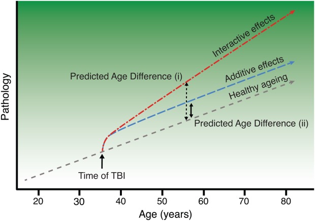
Objective: The long-term effects of traumatic brain injury (TBI) can resemble observed in normal ageing, suggesting that TBI may accelerate the ageing process. We investigate this using a neuroimaging model that predicts brain age in healthy individuals and then apply it to TBI patients. We define individuals' differences in chronological and predicted structural "brain age," and test whether TBI produces progressive atrophy and how this relates to cognitive function.

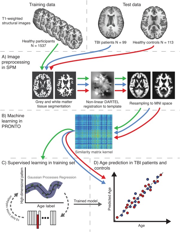
Methods: A predictive model of normal ageing was defined using machine learning in 1,537 healthy individuals, based on magnetic resonance imaging-derived estimates of gray matter (GM) and white matter (WM). This ageing model was then applied to test 99 TBI patients and 113 healthy controls to estimate brain age.

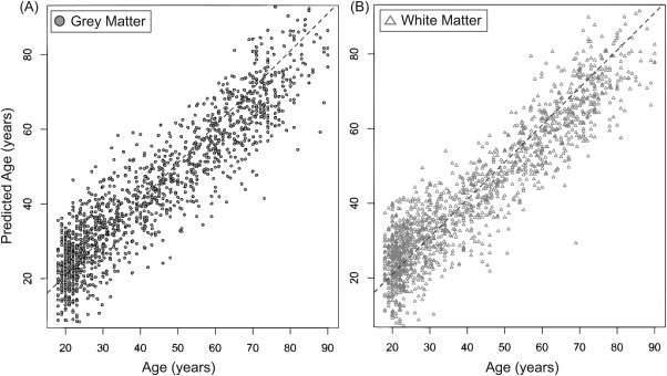
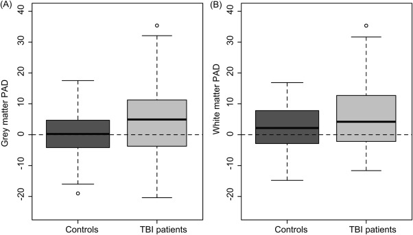
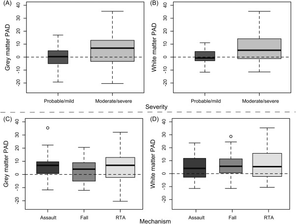
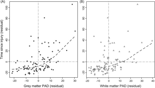
Results: The initial model accurately predicted age in healthy individuals (r = 0.92). TBI brains were estimated to be "older," with a mean predicted age difference (PAD) between chronological and estimated brain age of 4.66 years (±10.8) for GM and 5.97 years (±11.22) for WM. This PAD predicted cognitive impairment and correlated strongly with the time since TBI, indicating that brain tissue loss increases throughout the chronic postinjury phase.

Interpretation: TBI patients' brains were estimated to be older than their chronological age. This discrepancy increases with time since injury, suggesting that TBI accelerates the rate of brain atrophy. This may be an important factor in the increased susceptibility in TBI patients for dementia and other age-associated conditions, motivating further research into the age-like effects of brain injury and other neurological diseases.


Figures

Figure 1
Figure 1
Model of premature brain ageing in traumatic brain injury. Illustration of the conceptual framework for the investigation of brain age in traumatic brain injury (TBI). The short-dashed line represents the trajectory of healthy ageing as age (x-axis) increases, against a background gradient of increasing susceptibility to age-related pathology (y-axis), such as cognitive decline and dementia. Occurrence of TBI is indicated (black arrow), with acute pathology causing an immediate departure from a healthy brain state. Two alternative brain ageing trajectories post-TBI are shown. The long-dashed "additive effects" line depicts a trajectory assuming a one-off hit, with damage leading to the patient's brain structure resembling an older individual, followed by a normal rate of subsequent ageing. The dash–dot "interactive effects" line represents an accelerated rate of brain atrophy caused by TBI and an interaction with normal ageing processes, with the discrepancy between normal ageing and pathological changes increasing the greater the time since injury (TSI). Comparing predicted age difference (PAD) scores (i; dashed black line) and (ii; solid black line) illustrates how a greater PAD score would be expected under the interactive effects model with accelerating atrophy (i), compared to the added effects model (ii), at equivalent TSI (figure adapted from Smith and colleagues2). [Color figure can be viewed in the online issue, which is available at http://wileyonlinelibrary.com.]
Figure 2
Figure 2
Overview of the study methods. Study data comprised 2 sets, training and test. The training set used structural magnetic resonance imaging from 1,537 healthy individuals from multiple cohorts, whereas the test set included 2 groups, 99 traumatic brain injury (TBI) patients and 113 healthy controls, all scanned on the same scanner. (A) Conventional Statistical Parametric Mapping (SPM) structural preprocessing pipeline was used to generate gray and white matter maps, normalized to Montreal Neurological Institute (MNI) space and modulated to retain data relating to brain size. (B) Separately for gray and white matter, all 1,749 data sets were converted to a kernel matrix based on voxelwise similarity using Pronto. (C) The training data only were run through a supervised learning stage where a Gaussian Processes Regression (GPR) machine was trained to recognize patterns of imaging data that matched a given age label. To assess model accuracy, 10-fold cross-validation was conducted where 10% of samples were excluded from the training step and the ages of these samples were estimated. This was iterated 9 further times to generate age predictions on all samples. (D) The trained GPR model was then applied to the 2 test data sets, to assess accuracy of the model on healthy controls and then predict brain age of TBI patients. [Color figure can be viewed in the online issue, which is available at http://wileyonlinelibrary.com.]
Figure 3
Figure 3
Machine learning model provides accurate age prediction in healthy training set. Predicted age for each healthy individual in the training set (n * 1,537) is shown, derived by running 10-fold cross-validation on the Gaussian Processes Regression model. (A) Chronological age (x-axis) is plotted against predicted age (y-axis), for gray matter (dark gray circles). (B) Chronological age and predicted age for white matter (light gray triangles). Diagonal dashed line represents the line of identity (x * y).
Figure 4
Figure 4
Traumatic brain injury (TBI) patients show increased predicted age difference (PAD) score for gray matter and white matter. Boxplots of PAD score are shown, calculated by subtracting chronological age from predicted age for the test data sets of 99 TBI patients and 113 healthy controls. (A) PAD scores derived from the gray matter model showing a significant increase in TBI patients. (B) PAD scores from white matter also show an increase in TBI patients.
Figure 5
Figure 5
Gray matter (GM) and white matter (WM) predicted age difference (PAD) score, stratified by injury severity and injury mechanism. Boxplots of PAD score in the traumatic brain injury (TBI) patient group are shown, stratified by clinical characteristics. (A) GM PAD score distributions for each Mayo classification: probable/mild, moderate/severe, indicating that brain age is only increased in moderate/severe patients, not in mild TBI. (B) Mayo classification for WM. (C) GM PAD score by mechanism of injury (assault, fall, road–traffic accident [RTA]), indicating that similar levels of increased brain ageing occur independent of mechanism of injury. (D) Mechanism of injury for WM.
Figure 6
Figure 6
Gray matter and white matter predicted age difference (PAD) score increases with greater time since injury (TSI). Scatterplots depicting the relationship between PAD score (x-axis) and TSI (y-axis) are shown. Plotted values are the residuals derived from a linear regression with PAD score or TSI, regressing out chronological age. (A) Gray matter (dark gray circles) PAD scores, with dashed lines representing the locally weighted scatterplot smoothing (lowess) line calculated (dashed gray line). B) White matter (light gray triangles) PAD scores with lowess line (dashed light gray line). Both analyses were conducted after the removal of 3 outliers, identified based on having a TSI of ±2 standard deviations from the mean.

References

    1. Smith DH, Chen XH, Pierce JES. Progressive atrophy and neuron death for one year following brain trauma in the rat. J Neurotrauma. 1997;14:715–727. - PubMed
    1. Smith DH, Johnson VE, Stewart W. Chronic neuropathologies of single and repetitive TBI: Substrates of dementia? Nat Rev Neurol. 2013;9:211–221. - PMC - PubMed
    1. Moretti L, Cristofori I, Weaver SM. Cognitive decline in older adults with a history of traumatic brain injury. Lancet Neurol. 2012;11:1103–1112. - PubMed
    1. Bazarian JJ, Cernak I, Noble-Haeusslein L. Long-term neurologic outcomes after traumatic brain injury. J Head Trauma Rehabil. 2009;24:439–451. - PubMed
    1. Hedden T, Gabrieli JDE. Insights into the ageing mind: a view from cognitive neuroscience. Nat Rev Neurosci. 2004;5:87–96. - PubMed

Publication types