Case Reports
. 2015 Jan 27:8:59.
doi: 10.1186/s13071-015-0655-2.

New zoonotic cases of Onchocerca dewittei japonica (Nematoda: Onchocercidae) in Honshu, Japan

Affiliations
Case Reports

New zoonotic cases of Onchocerca dewittei japonica (Nematoda: Onchocercidae) in Honshu, Japan

Shigehiko Uni et al. Parasit Vectors. .

Abstract

Background: Zoonotic infections with Onchocerca species are uncommon, and to date only 25 clinical cases have been reported worldwide. In Japan, five previous zoonotic infections were concentrated in Oita, Kyushu (the southern island), with one previous case in Hiroshima in the western part of Honshu (the main island). The causative agent in Japan was identified as Onchocerca dewittei japonica Uni, Bain & Takaoka, 2001 from Japanese wild boars (Sus scrofa leucomystax Temminck, 1842). Here we report two infections caused by a female and male O. dewittei japonica, respectively, among residents of Hiroshima and Shimane Prefectures in the western part of Honshu.

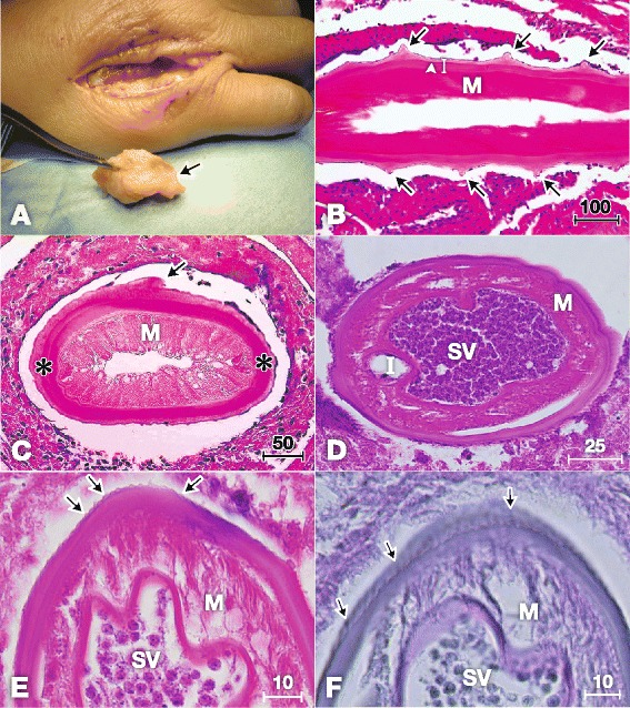
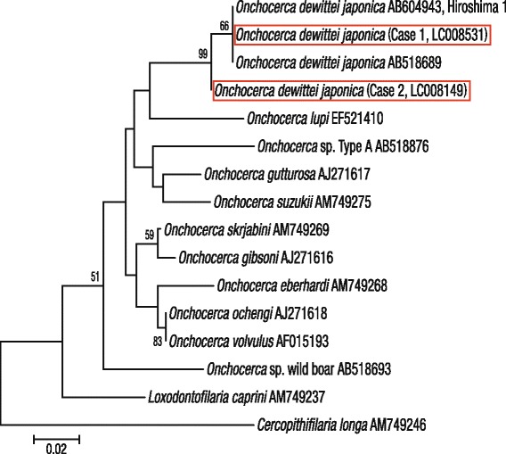
Methods: In both cases, nodules were surgically removed. The parasites in nodules were identified on the basis of their histopathological characteristics. Identification was confirmed by sequencing the mitochondrial cytochrome c oxidase subunit 1 (cox1) gene from worms in the tissues used in the histological preparations.

Results: Case 1 was a 61-year-old woman from Hiroshima Prefecture who complained of a painful subcutaneous nodule on the back of her right hand. The causative agent was identified as a female O. dewittei japonica owing to transverse ridges on the cuticle and molecular analysis. Case 2 was a 78-year-old woman from Shimane Prefecture who had a painful nodule in the left temporal region. Histopathological characteristics and cox1 sequencing of the worm indicated that the causative agent was a male O. dewittei japonica.

Conclusions: For Cases 1 and 2, we diagnosed the causative agents as a female and male O. dewittei japonica, respectively. These findings indicate the spread of a zoonosis caused by O. dewittei japonica in the western part of Honshu, where wild boars have recently extended their habitats because of decreased annual snowfall, unused rice fields and a decline in the number of hunters in Japan. The O. dewittei japonica infection rate among wild boars was reported as 78% in Shimane Prefecture, in the western part of Honshu. Therefore, in the near future, zoonotic onchocercosis is likely to occur in Honshu as well as Kyushu, where wild boars, blackfly vectors and humans share the same habitat.


Figures

Figure 1
Figure 1
Histopathological characteristics of Onchocerca dewittei japonica in nodules excised from Cases 1 in Hiroshima (A–C) and 2 in Shimane (D–F). A) Mass (arrow, 1 × 3 cm) excised from the back of the right hand. B) Longitudinal section of the female O. dewittei japonica. Arrows, triangular transverse ridges; arrowhead, middle layer of the cuticle without the inner striae; white vertical line, cuticle; M, muscle layer. HE staining. C) Slightly oblique transverse section of the female worm showing the thick cuticle. Arrow, elevation of the cuticle, indicating the transverse ridge; asterisks, lateral chords; M, muscle layer. HE staining. D) Transverse section of the male O. dewittei japonica. I, intestine; M, muscle layer; SV, seminal vesicle with spermatozoids. HE staining. E) Enlarged transverse section. Arrows, small ridges on the cuticle; M, muscle layer; SV, seminal vesicle. HE staining. F) Slightly oblique transverse section. Arrows, small longitudinal ridges; M, muscle layer; SV, seminal vesicle. PAS staining. Unit of bars, μm.
Figure 2
Figure 2
Molecular identification of the causative agents ( Onchocerca dewittei japonica ) of Cases 1 and 2 (red rectangles). Phylogeny of Onchocerca spp. based on GenBank sequences of the cox1 using the neighbour-joining method. The numbers at the nodes indicate the bootstrap confidence values on the basis of 500 replicates, where values >50% are shown. Bar, the number of changes inferred to have occurred along each branch.
Figure 3
Figure 3
Geographic distribution of human zoonotic onchocercosis in Japan. Case 1 (*) in the current study and one previous case in Hiroshima; Case 2 (*) in Shimane; five previous cases reported in Oita, Kyushu. The green areas indicate habitat of wild boars in Japan in 2003 based on ‘The Sixth National Survey on the Natural Environmental Report of the Distributional Survey of Japanese Animals (Mammals)’ [25].

References

    1. Orihel TC, Eberhard ML. Zoonotic filariasis. Clin Microbiol Rev. 1998;11:366–81. - PMC - PubMed
    1. Otranto D, Eberhard ML. Zoonotic helminths affecting the human eye. Parasit Vectors. 2011;4:41. doi: 10.1186/1756-3305-4-41. - DOI - PMC - PubMed
    1. Colwell DD, Dantas-Torres F, Otranto D. Vector-borne parasitic zoonoses: emerging scenarios and new perspectives. Vet Parasitol. 2011;182:14–21. doi: 10.1016/j.vetpar.2011.07.012. - DOI - PubMed
    1. Takaoka H, Fukuda M, Otsuka Y, Aoki C, Uni S, Bain O. Blackfly vectors of zoonotic onchocerciasis in Japan. Med Vet Entomol. 2012;26:372–8. doi: 10.1111/j.1365-2915.2012.01023.x. - DOI - PubMed
    1. Uni S, Boda T, Daisaku K, Ikura Y, Maruyama H, Hasegawa H, et al. Zoonotic filariasis caused by Onchocerca dewittei japonica in a resident of Hiroshima Prefecture, Honshu, Japan. Parasitol Int. 2010;59:477–80. doi: 10.1016/j.parint.2010.05.006. - DOI - PubMed

Publication types