. 2015 Feb 10;112(6):E516-25.
doi: 10.1073/pnas.1424651112. Epub 2015 Jan 26.

Membranes serve as allosteric activators of phospholipase A2, enabling it to extract, bind, and hydrolyze phospholipid substrates

Affiliations

Membranes serve as allosteric activators of phospholipase A2, enabling it to extract, bind, and hydrolyze phospholipid substrates

Varnavas D Mouchlis et al. Proc Natl Acad Sci U S A. .

Abstract

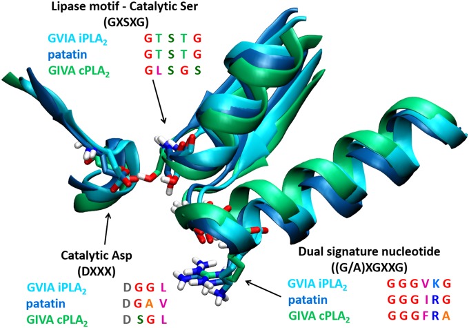
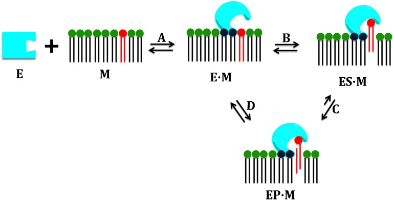
Defining the molecular details and consequences of the association of water-soluble proteins with membranes is fundamental to understanding protein-lipid interactions and membrane functioning. Phospholipase A2 (PLA2) enzymes, which catalyze the hydrolysis of phospholipid substrates that compose the membrane bilayers, provide the ideal system for studying protein-lipid interactions. Our study focuses on understanding the catalytic cycle of two different human PLA2s: the cytosolic Group IVA cPLA2 and calcium-independent Group VIA iPLA2. Computer-aided techniques guided by deuterium exchange mass spectrometry data, were used to create structural complexes of each enzyme with a single phospholipid substrate molecule, whereas the substrate extraction process was studied using steered molecular dynamics simulations. Molecular dynamic simulations of the enzyme-substrate-membrane systems revealed important information about the mechanisms by which these enzymes associate with the membrane and then extract and bind their phospholipid substrate. Our data support the hypothesis that the membrane acts as an allosteric ligand that binds at the allosteric site of the enzyme's interfacial surface, shifting its conformation from a closed (inactive) state in water to an open (active) state at the membrane interface.

Keywords: DXMS; GIVA cPLA2; GVIA iPLA2; MD simulations; PAPC.


Conflict of interest statement

The authors declare no conflict of interest.

Figures

Fig. 1.
Fig. 1.
Sequence and structure alignment of GVIA iPLA2 (cyan), patatin (blue), and GIVA cPLA2 (green).
Fig. 2.
Fig. 2.
Defining the binding pockets using SiteMap and predicting the PAPC–enzyme complexes using IFD for GIVA cPLA2 and GVIA iPLA2. (A) The binding pocket of GIVA cPLA2, and (B) the binding pocket of GVIA iPLA2: hydrophobic regions are highlighted in magenta and hydrophilic in light blue. (C) PAPC-GIVA cPLA2 complex, and (D) PAPC-GVIA iPLA2 complex predicted by IFD.
Fig. 3.
Fig. 3.
Predicting the PAPC–enzyme complexes using SMD simulations for GIVA cPLA2 and GVIA iPLA2. (A) The system of GIVA cPLA2 and PAPC embedded in a POPC membrane used for SMD simulations, in which the PAPC was pulled into the binding pocket in the direction depicted by the black arrow. (B) The system of GVIA iPLA2 and PAPC embedded in a POPC membrane used for SMD simulations, in which the PAPC was pulled into the binding pocket in the direction depicted by the black arrow. (C) PAPC–GIVA cPLA2 complex predicted by SMD (Movie S1). (D) PAPC–GVIA iPLA2 complex predicted by SMD (Movie S2).
Fig. 4.
Fig. 4.
PAPC binding mode, number of H-bonds, and volume of the binding pocket for GIVA cPLA2 and GVIA iPLA2 after 300-ns MD simulation. (A) Binding mode of PAPC in the GIVA cPLA2 binding pocket (Movie S3). (B) Binding mode of PAPC in the GVIA iPLA2 binding pocket (Movie S4). (C) Average number of H-Bonds of the PAPC with the residues of the binding pocket for each enzyme during the simulation. (D) Volume of the binding pocket of each enzyme during the simulation (Movies S7 and S8).
Fig. 5.
Fig. 5.
The dynamic nature of the GVIA iPLA2 binding pocket. Volume of the binding pocket during the MD simulation of (A) the GVIA iPLA2-PAPC complex, (B) the open state without PAPC in the pocket (although PAPC is superimposed for visual convenience) in the presence of a membrane patch containing POPC, (C) the open state without PAPC (although PAPC is superimposed for visual convenience) in the pocket in the presence of water. (D) Volume of the binding pocket during the MD simulation (A, cyan; B, brown; and C, blue; Movie S9).
Fig. 6.
Fig. 6.
Representation of the catalytic cycle of phospholipases A2. (A) Membrane association shifts the enzyme conformation from closed to open. (B) Extraction and binding of a single phospholipid molecule. (C) Hydrolysis of the bound phospholipid molecule. (D). Diffusion of the products into the membrane.

References

    1. Dennis EA, Cao J, Hsu YH, Magrioti V, Kokotos G. Phospholipase A2 enzymes: Physical structure, biological function, disease implication, chemical inhibition, and therapeutic intervention. Chem Rev. 2011;111(10):6130–6185. - PMC - PubMed
    1. Buczynski MW, Dumlao DS, Dennis EA. Thematic Review Series: Proteomics. An integrated omics analysis of eicosanoid biology. J Lipid Res. 2009;50(6):1015–1038. - PMC - PubMed
    1. Pniewska E, Pawliczak R. The involvement of phospholipases A2 in asthma and chronic obstructive pulmonary disease. Mediators Inflamm. 2013;2013:793505. - PMC - PubMed
    1. Masuda S, et al. Various secretory phospholipase A2 enzymes are expressed in rheumatoid arthritis and augment prostaglandin production in cultured synovial cells. FEBS J. 2005;272(3):655–672. - PubMed
    1. Rosenson RS, Hurt-Camejo E. Phospholipase A2 enzymes and the risk of atherosclerosis. Eur Heart J. 2012;33(23):2899–2909. - PubMed

Publication types