. 2015 Feb 10;112(6):1749-54.
doi: 10.1073/pnas.1416656112. Epub 2015 Jan 26.

Src phosphorylation converts Mdm2 from a ubiquitinating to a neddylating E3 ligase

Affiliations

Src phosphorylation converts Mdm2 from a ubiquitinating to a neddylating E3 ligase

Christopher N Batuello et al. Proc Natl Acad Sci U S A. .

Abstract

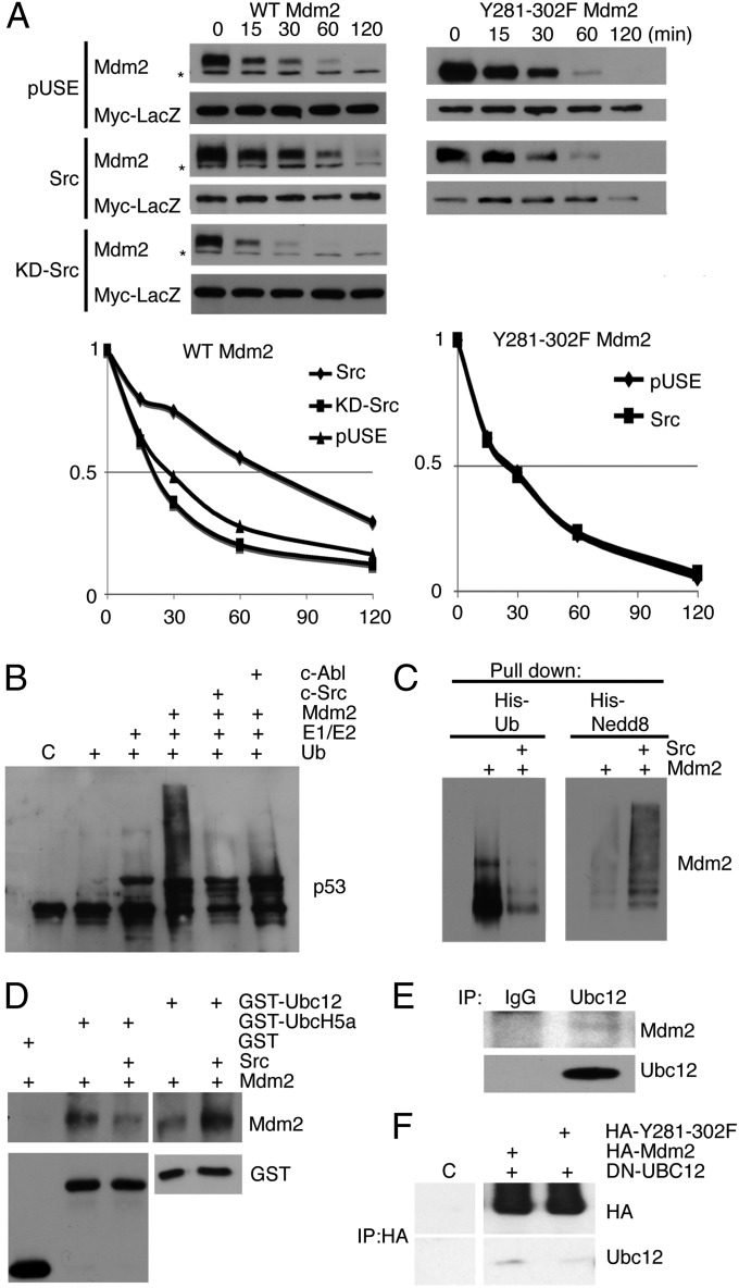
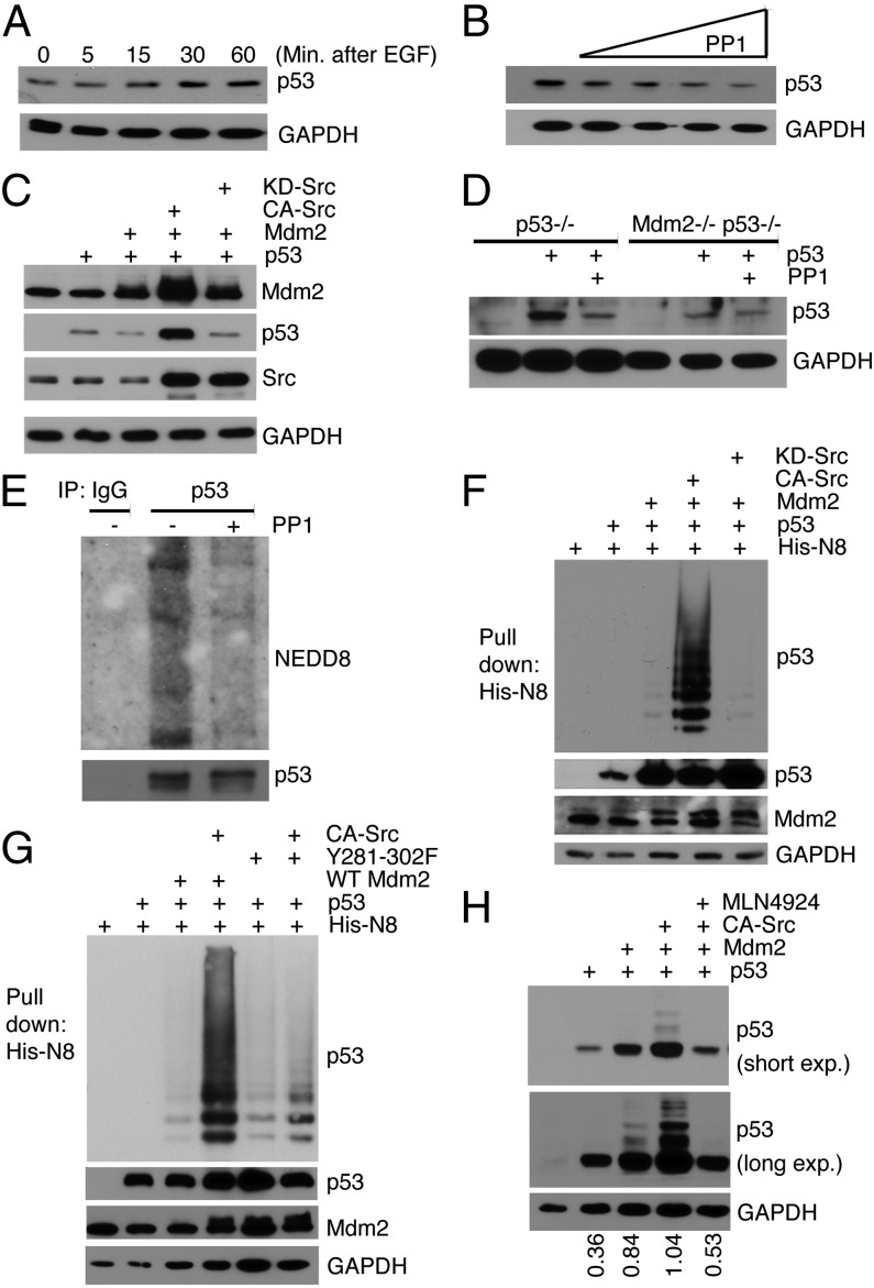
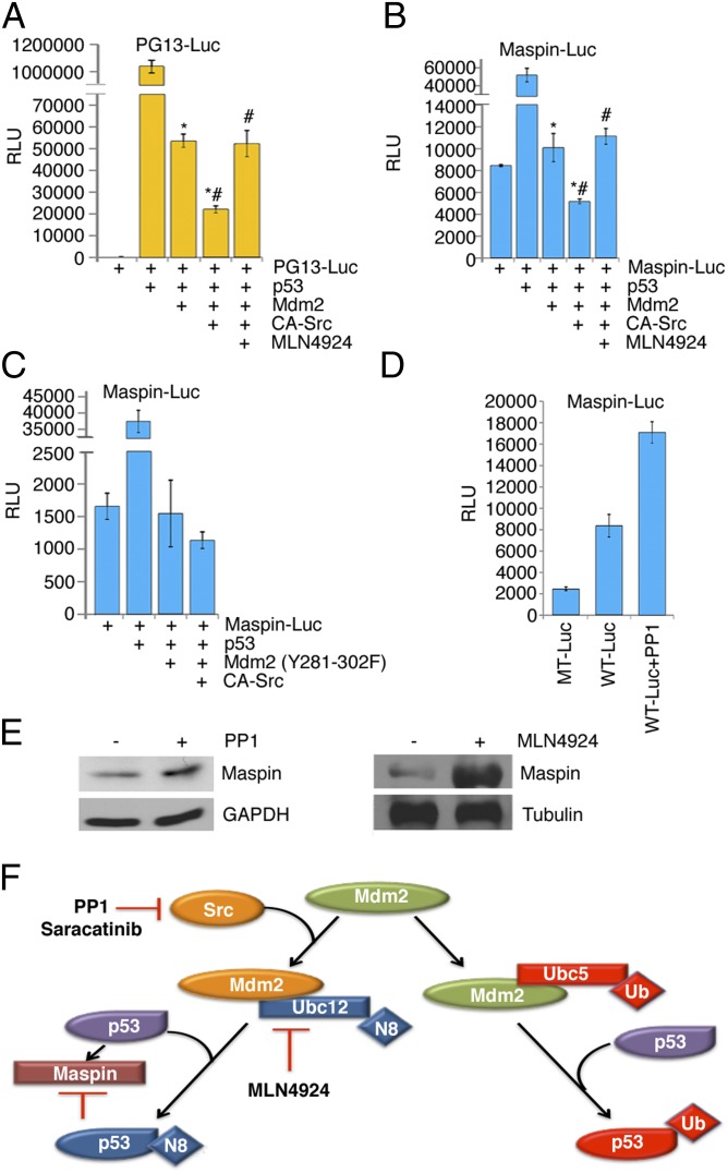
Murine double minute-2 protein (Mdm2) is a multifaceted phosphorylated protein that plays a role in regulating numerous proteins including the tumor suppressor protein p53. Mdm2 binds to and is involved in conjugating either ubiquitin or Nedd8 (Neural precursor cell expressed, developmentally down-regulated 8) to p53. Although regulation of the E3 ubiquitin activity of Mdm2 has been investigated, regulation of the neddylating activity of Mdm2 remains to be defined. Here we show that activated c-Src kinase phosphorylates Y281 and Y302 of Mdm2, resulting in an increase in Mdm2 stability and its association with Ubc12, the E2 enzyme of the neddylating complex. Mdm2-dependent Nedd8 conjugation of p53 results in transcriptionally inactive p53, a process that is reversed with a small molecule inhibitor to either Src or Ubc12. Thus, our studies reveal how Mdm2 may neutralize and elevate p53 in actively proliferating cells and also provides a rationale for using therapies that target the Nedd8 pathway in wild-type p53 tumors.

Keywords: Mdm2; Nedd8; Src.


Conflict of interest statement

The authors declare no conflict of interest.

Figures

Fig. 1.
Fig. 1.
Mdm2 and c-Src form a complex in vitro and in vivo. (A) Schematic of Mdm2 is shown (Top). His-tagged Mdm2 and GST-tagged SH3Src from a nickel pull-down of His-Mdm2, His-Mdm2 SH3 domain mutant 1, and His-Mdm2 SH3 domain mutant 2 were immunoblotted for GST and His antibodies. Input is GST-SH3Src. (B) Immunoprecipitations of endogenous Mdm2 from 293T (Left) or c-Src from MCF7 and U87 cell extracts (Center and Right) were probed with Src and Mdm2 antibodies. (C) Mdm2 and c-Src were detected by immunoblot of cytoplasmic and nuclear extracts from MCF7 cells. Detection of tubulin and PARP indicate extract purity.
Fig. 2.
Fig. 2.
c-Src phosphorylates Mdm2 at tyrosines 281 and 302. (A) The total and the defined tyrosine phosphorylation sites (P) in Mdm2 are illustrated. (B) An in vitro phosphorylation reaction of Mdm2 (p-Mdm2) by c-Src or c-Abl using 32P was detected by autoradiography. Autophosphorylation of c-Src and c-Abl, along with c-Abl phosphorylation of Mdm2, served as internal controls. (C) Immunoblot of in vitro phosphorylation reactions with Src and Mdm2 was probed with the 4G10 antibody to detect phospho-tyrosines (pTyr) (Right). (Left) Immunoblot of the input probed with SMP14. (D) HA-Mdm2 was immunopurified from H1299 cells that have been transiently transfected with HA-Mdm2 and KD-Src or CA-Src. Immunoblots were probed with anti-HA antibodies or the p-Tyr antibody, 4G10, to show tyrosine phosphorylation of Mdm2. Immunoblot (Bottom) was probed with Src to show expression in lysates. (E) Mdm2 was immunoprecipitated from MCF7 extracts after treatment with 10 μM PP1 or DMSO for 16 h and immunoblotted for p-Tyr and Mdm2. (F) In vitro phosphorylation reactions were performed with Src on His-tagged 90–383 Mdm2 and the Y281-302F double mutant.
Fig. 3.
Fig. 3.
c-Src phosphorylation results in an enhancement of Mdm2 protein levels. (A) Immunoblot analysis was performed with H1229 cells overexpressing Mdm2 that had been treated with increasing concentrations (0.5, 1, 5, 10 μg) of CA-Src. (B) Cellular extracts of H1299 cells ectopically expressing Mdm2 alone or with CA-Src or KD-Src were immunoblotted for Mdm2, c-Src, and GAPDH. (C) MCF7 cells were serum starved for 48 h and then treated with 200 ng/mL EGF for the time indicated. Endogenous Mdm2, p-Src, and GAPDH (for loading) were analyzed by immunoblot. The asterisk indicates a nonspecific band. (D) MCF7 cells were serum starved for 48 h and then pretreated with either DMSO or PP1 (10 μM) for 1 h before the addition of EGF (200 ng/mL). EGF treatment of 1 h was followed by immunoblot analysis of Mdm2 and GAPDH. The asterisk indicates a nonspecific band. (E) Mdm2 levels (and GAPDH as control) in MCF7 cells pretreated with saracatanib or DMSO overnight followed by EGF for 1 h were detected by immunoblotting. The asterisk indicates a nonspecific band.
Fig. 4.
Fig. 4.
c-Src expression increases the half-life of Mdm2 through inhibition of its E3 ubiquitination activity. (A) H1299 cells were transfected with Mdm2 (Left) or Y281-302F Mdm2 (Right) and CA-Src, KD-Src, or empty vector (pUSE) in the presence of Myc-LacZ vector. Twenty-four hours posttransfection, the cells were treated with 50 μg/mL cycloheximide (CHX) and harvested at different points, as indicated. The cell lysates were immunoblotted with anti-Mdm2 and anti-Myc antibodies, as indicated (Top). The density of Mdm2 in each lane was quantified against the level of Myc-LacZ and plotted in a graph (Bottom). The asterisk indicates a nonspecific band. (B) An in vitro ubiquitination assay was performed with recombinant p53, using Mdm2 or Mdm2 phosphorylated by c-Src/c-Abl. Immunoblot was probed for p53 (D01). (C) A His pull-down assay was used to determine whether Mdm2 was either His-ubiquitinated (Left) or His-neddylated (Right) in the presence of CA-Src. Immunoblots were used to detect Mdm2. (D) A GST-pull-down of GST, GST-Ubc5, or GST-Ubc12 was used to determine whether Mdm2 or c-Src-phosphorylated Mdm2 would preferentially complex to an E2. Mdm2 and GST were detected by immunoblot. (E) Cellular extracts from MCF7 cells were used to immunoprecipitate Ubc12 or isotype control. Mdm2 and Ubc12 were detected by immunoblot. (F) Immunoblots detected Ubc12 and HA, from immunoprecipitations of HA from 239T cells transiently transfected with combinations of HA-Mdm2, HA-Y281-302F Mdm2, and DN-Ubc12.
Fig. 5.
Fig. 5.
p53 protein levels are regulated by c-Src-phosphorylated Mdm2. (A) MCF7 cells were serum starved for 48 h and incubated with EGF over a time course of 1 h. Extracts were immunoblotted for p53 and GAPDH. (B) MCF7 cells were treated with increasing concentrations of PP1 (1, 5, 10, 15, 20 μM) for 16 h. Cellular extracts were immunoblotted for p53 and GAPDH. (C) Lysates from H1299 cells overexpressing combinations of p53, Mdm2, CA-Src, or KD-Src were immunoblotted for Mdm2, p53, Src, and GAPDH. (D) Cellular extracts from p53−/− or p53−/− Mdm2−/− MEFs that had been transiently transfected with p53 and treated with PP1 (10 μM) or DMSO were immunoblotted for p53 and GAPDH. (E) Immunoprecipitations of p53 and IgG control from MCF7 cellular extracts were used to detect endogenous Nedd8 and p53 in immunoblots. Cells were either treated with PP1 (10 μM) or DMSO for 16 h. (F) p53 was detected by immunoblot from a His-Nedd8 pull-down assay conducted in H1299 cells expressing His-Nedd8, p53, WT-Mdm2, CA-Src, or KD-Src. To verify expression, lysates were subjected to immunoblot analysis, as indicated. (G) His-Nedd8 pull-down was performed as in F, but with the use of Mdm2-Y281-302F, as indicated. (H) H1299 cells were transfected with p53, Mdm2, and CA-Src 24 h before treatment with either DMSO or 0.3 μM of the Nedd8-activating enzyme inhibitor MLN4924 for 16 h. Immunoblot detected p53 and GAPDH and relative amounts of p53 were determined.
Fig. 6.
Fig. 6.
Increased p53 levels do not translate to increased transcriptional activity. (A) PG13-Luc activity was assayed using H1299 cells transfected with Myc-LacZ, p53, Mdm2, and CA-Src. Twenty-four hours posttransfection, cells were treated with DMSO or 0.3 μM of the Nedd8-activating enzyme inhibitor MLN4924 for 16 h. (B) H1299 cells were treated the same as in A, except luciferase was assayed using Maspin-Luc and Myc-LacZ. (C) maspin-luciferase activity was examined in H1299 cells transfected with p53, Mdm2 Y281-302F, and CA-Src. (D) Luciferase assay of MCF7 cells transfected with either Maspin-Luc or Mutant Maspin-Luc (MT) and Myc-LacZ was conducted. Twenty-four hours after transfection, cells were treated with DMSO or 10 μM PP1 for 16 h. (AD) Y axis measurements are relative luciferase units (RLU). The RLU were calculated from the ratio of luciferase/β-gal activity. Error bars represent SD of three separate experiments. *,#P < 0.05. (E) MCF7 cells were treated with DMSO or 10 μM PP1 and subjected to immunoblotting for Maspin and GAPDH. (F) In response to activated Src, Mdm2 is phosphorylated and selectively binds to Ubc12 to function as a neddylating enzyme. The respective inhibitors to the pathway are denoted.

Similar articles

Cited by

References

    1. Yeatman TJ. A renaissance for SRC. Nat Rev Cancer. 2004;4(6):470–480. - PubMed
    1. Shalloway D, Coussens PM, Yaciuk P. Overexpression of the c-src protein does not induce transformation of NIH 3T3 cells. Proc Natl Acad Sci USA. 1984;81(22):7071–7075. - PMC - PubMed
    1. Biscardi JS, Ishizawar RC, Silva CM, Parsons SJ. Tyrosine kinase signalling in breast cancer: Epidermal growth factor receptor and c-Src interactions in breast cancer. Breast Cancer Res. 2000;2(3):203–210. - PMC - PubMed
    1. Manning BD, Cantley LC. AKT/PKB signaling: Navigating downstream. Cell. 2007;129(7):1261–1274. - PMC - PubMed
    1. Mayo LD, Donner DB. A phosphatidylinositol 3-kinase/Akt pathway promotes translocation of Mdm2 from the cytoplasm to the nucleus. Proc Natl Acad Sci USA. 2001;98(20):11598–11603. - PMC - PubMed

Publication types