. 2015 Feb 10;112(6):1761-6.
doi: 10.1073/pnas.1413185112. Epub 2015 Jan 26.

Direct inhibition of oncogenic KRAS by hydrocarbon-stapled SOS1 helices

Affiliations

Direct inhibition of oncogenic KRAS by hydrocarbon-stapled SOS1 helices

Elizaveta S Leshchiner et al. Proc Natl Acad Sci U S A. .

Abstract

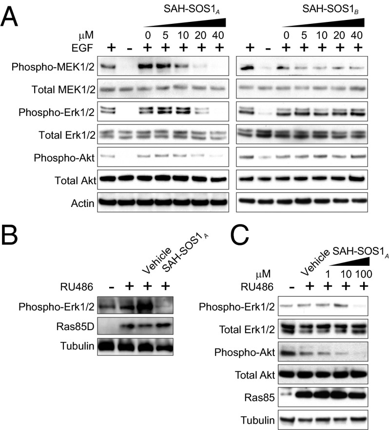
Activating mutations in the Kirsten rat sarcoma viral oncogene homolog (KRAS) underlie the pathogenesis and chemoresistance of ∼ 30% of all human tumors, yet the development of high-affinity inhibitors that target the broad range of KRAS mutants remains a formidable challenge. Here, we report the development and validation of stabilized alpha helices of son of sevenless 1 (SAH-SOS1) as prototype therapeutics that directly inhibit wild-type and mutant forms of KRAS. SAH-SOS1 peptides bound in a sequence-specific manner to KRAS and its mutants, and dose-responsively blocked nucleotide association. Importantly, this functional binding activity correlated with SAH-SOS1 cytotoxicity in cancer cells expressing wild-type or mutant forms of KRAS. The mechanism of action of SAH-SOS1 peptides was demonstrated by sequence-specific down-regulation of the ERK-MAP kinase phosphosignaling cascade in KRAS-driven cancer cells and in a Drosophila melanogaster model of Ras85D(V12) activation. These studies provide evidence for the potential utility of SAH-SOS1 peptides in neutralizing oncogenic KRAS in human cancer.

Keywords: RAS; SOS1; cancer; inhibitor; stapled peptide.


Conflict of interest statement

Conflict of interest statement: L.D.W. is a scientific advisory board member and consultant for Aileron Therapeutics.

Figures

Fig. 1.
Fig. 1.
Design and KRAS binding activity of SAH-SOS1 peptides. (A) The crystal structure of KRAS (red) in complex with its guanidine exchange factor SOS1 (blue) revealed a binding interaction between the indicated SOS1 α-helix (cyan) and KRAS (PDB ID code 1NVU). SAH-SOS1 peptides were generated by inserting all-hydrocarbon staples at positions A (green) and B (orange) into a SOS1 peptide spanning amino acids 929–944 and bearing an N-terminal Arg-Arg tag to optimize cellular penetrance. (B) Fluorescence polarization binding analysis of FITC-labeled SAH-SOS1 peptides and recombinant KRAS proteins, including wild-type and mutant variants. Data are mean ± SEM for experiments performed in technical triplicate and are representative of at least three biological replicates performed with independent preparations of recombinant KRAS proteins. (C) Table of EC50 values for the binding interactions between SAH-SOS1 peptides and the individual KRAS proteins.
Fig. 2.
Fig. 2.
NMR analysis of SAH-SOS1A/KRAS interaction. (A) 1H-15N HSQC spectrum of uniformly 15N-labeled GDP-loaded WT KRAS in the absence (red) or presence of SAH-SOS1A (blue). (B) Chemical shift changes (significance threshold > 0.01 ppm) are plotted as a function of WT KRAS residue number. (C) SAH-SOS1A was docked onto KRAS (starting structural model PDB ID code 1NVU; HADDOCK software) based on HSQC data. The calculated model structure depicts the SOS1 helix engaging the SOS1-binding pocket of KRAS.
Fig. 3.
Fig. 3.
SAH-SOS1A inhibits nucleotide association with wild-type and mutant KRAS. (A and B) The indicated fluorescent nucleotide analogs were incubated with wild-type (A) or G12D mutant (B) KRAS protein and the increase in fluorescence was monitored over time (red). Coincubation with unlabeled nucleotide served as a negative control (black). A serial dilution of SAH-SOS1 peptides were coincubated with KRAS and fluorescent nucleotide at the indicated doses to monitor for effects on nucleotide association. Data are mean ± SEM for experiments performed in at least technical triplicates and are representative of two biological replicates performed with independent preparations of recombinant KRAS proteins.
Fig. 4.
Fig. 4.
SAH-SOS1A cytotoxicity correlates with KRAS binding activity. (A) SAH-SOS1A, but not SAH-SOS1B, dose-responsively inhibits the viability of Panc 10.05 cells bearing the KRAS G12D mutation. Cells were treated with vehicle, SAH-SOS1A, or SAH-SOS1B, and cell viability was measured at 24 h by CellTiterGlo assay. Data are mean ± SEM for experiments performed in at least duplicate and representative of at least two biological replicates performed with independent cancer cell cultures. (B) A panel of SAH-SOS1A mutant peptides was generated for structure activity relationship studies. (C) Fluorescence polarization binding analysis of SAH-SOS1A peptides and recombinant KRAS G12D protein. Data are mean ± SEM for experiments performed in technical duplicate and representative of at least three biological replicates performed with independent preparations of recombinant KRAS protein. (D) Panc 10.05 cells were treated with the panel of SAH-SOS1A peptides and cell viability measured at 24 h by CellTiterGlo assay. Data are mean ± SEM for experiments performed in at least technical duplicates and are representative of at least two biological replicates performed with independent cancer cell cultures.
Fig. 5.
Fig. 5.
SAH-SOS1A inhibits phosphosignaling downstream of KRAS in vitro and in vivo. (A) Panc 10.05 cells were incubated with vehicle, SAH-SOS1A, or SAH-SOS1B at the indicated doses for 4 h, followed by 15-min stimulation with EGF. Cellular lysates were then electrophoresed and subjected to Western blot analysis by using antibodies to phospho- and total MEK1/2, ERK1/2, and Akt. (B) Vehicle (DMSO) or SAH-SOS1A (0.2 μL of 10 mM solution) was injected into the abdomens of D. melanogaster Ras85DV12/ActinGS (n = 5 per arm) after a 3-d period of RAS induction by RU486 treatment (150 μg/mL in 2 mL of fly food). Flies were collected 48 h after SAH-SOS1A peptide injection, and lysates were processed for electrophoresis and Western blot analysis by using anti-phospho-ERK1/2 antibody. (C) D. melanogaster Ras85DV12/ActinGS (n = 20 per arm) treated with vehicle or RU486 (150 μg/mL) alone, or cotreated with RU486 and SAH-SOS1A (1, 10, or 100 μM in 2 mL of fly food). After 4 d of oral treatment, tissue lysates were subjected to electrophoresis and Western blotting for phospho- and total ERK1/2 and Akt.

References

    1. Karnoub AE, Weinberg RA. Ras oncogenes: Split personalities. Nat Rev Mol Cell Biol. 2008;9(7):517–531. - PMC - PubMed
    1. Stephen AG, Esposito D, Bagni RK, McCormick F. Dragging ras back in the ring. Cancer Cell. 2014;25(3):272–281. - PubMed
    1. Hidalgo M. Pancreatic cancer. N Engl J Med. 2010;362(17):1605–1617. - PubMed
    1. Lim SM, et al. Therapeutic targeting of oncogenic K-Ras by a covalent catalytic site inhibitor. Angew Chem Int Ed Engl. 2014;53(1):199–204. - PMC - PubMed
    1. Ostrem JM, Peters U, Sos ML, Wells JA, Shokat KM. K-Ras(G12C) inhibitors allosterically control GTP affinity and effector interactions. Nature. 2013;503(7477):548–551. - PMC - PubMed

Publication types

MeSH terms

LinkOut - more resources