. 2012 Sep 15;7(26):2018-27.
doi: 10.3969/j.issn.1673-5374.2012.26.003.

Human umbilical cord blood-derived mesenchymal stem cells promote regeneration of crush-injured rat sciatic nerves

Affiliations

Human umbilical cord blood-derived mesenchymal stem cells promote regeneration of crush-injured rat sciatic nerves

Mi-Ae Sung et al. Neural Regen Res. .

Abstract

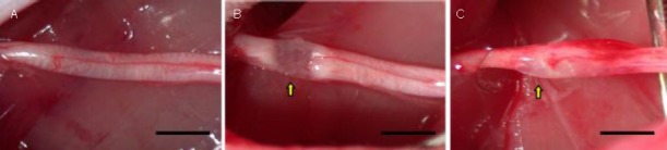
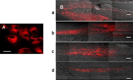
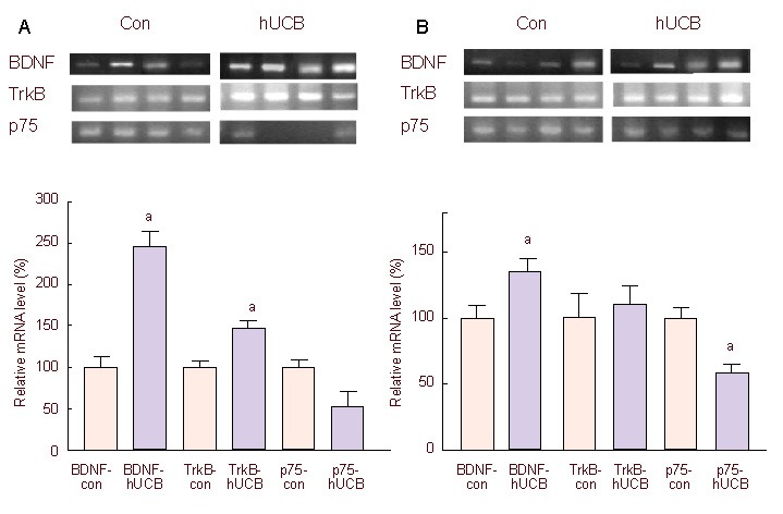
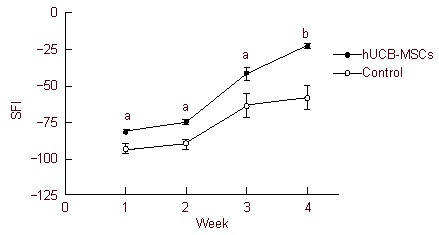
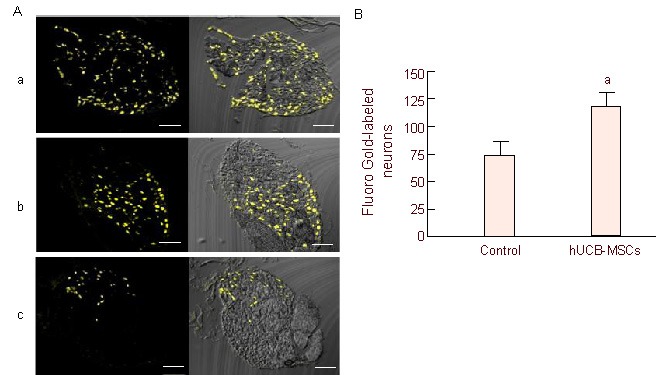
Several studies have demonstrated that human umbilical cord blood-derived mesenchymal stem cells can promote neural regeneration following brain injury. However, the therapeutic effects of human umbilical cord blood-derived mesenchymal stem cells in guiding peripheral nerve regeneration remain poorly understood. This study was designed to investigate the effects of human umbilical cord blood-derived mesenchymal stem cells on neural regeneration using a rat sciatic nerve crush injury model. Human umbilical cord blood-derived mesenchymal stem cells (1 × 10(6)) or a PBS control were injected into the crush-injured segment of the sciatic nerve. Four weeks after cell injection, brain-derived neurotrophic factor and tyrosine kinase receptor B mRNA expression at the lesion site was increased in comparison to control. Furthermore, sciatic function index, Fluoro Gold-labeled neuron counts and axon density were also significantly increased when compared with control. Our results indicate that human umbilical cord blood-derived mesenchymal stem cells promote the functional recovery of crush-injured sciatic nerves.

Keywords: Fluoro Gold; crush injury; human umbilical cord blood-derived mesenchymal stem cells; neural regeneration; peripheral nerve regeneration; regeneration; sciatic nerve; stem cells.


Conflict of interest statement

Conflicts of interest: None declared.

Figures

Figure 1
Figure 1
A rat model of sciatic nerve crush injury. (A) The normal sciatic nerve; (B) crush-injured sciatic nerve, nerve fibers were discontinuous with preservation of the epineurium; and (C) crush injured sciatic nerve after human umbilical cord blood-derived mesenchymal stem cell transplantation. Arrows indicate the lesion. Bars: 5 mm.
Figure 2
Figure 2
Confocal photomicrographs of PKH26-labeled (red fluorescence) human umbilical cord blood-derived mesenchymal stem cells (hUCB-MSCs) before and after transplantation. (A) PKH26-labeled hUCB-MSCs before transplantation, captured 2 hours after seeding on a glass slide. Bar: 20 μm. (B) PKH26-labeled hUCB-MSCs in the lesion site at 1 week (a), 2 weeks (b), 3 weeks (c), and 4 weeks after transplantation (d). Bars: 200 μm.
Figure 3
Figure 3
Relative mRNA expression of BDNF, TrkB and p75 in the sciatic nerve (A) and L4-6 DRGs (B), 5 days after injection of hUCB-MSCs (hUCB-MSCs group: hUCB) or PBS (control group: con). All mRNA was normalized to GAPDH. mRNA expression levels of BDNF, TrkB and p75 in the hUCB-MSCs group were relative to that of the control group. aP < 0.05, vs. control group. Statistical significance was tested by analysis of variance. Data are presented as mean ± SEM; n = 6. hUCB-MSCs: Human umbilical cord blood-derived mesenchymal stem cells; TrkB: tyrosine kinase receptor B; BDNF: brain-derived neurotrophic factor.
Figure 4
Figure 4
SFI value in rats with sciatic nerve injury. The SFI value of both groups was increased, however, SFI in the hUCB-MSCs group was significantly higher than the control group at all time points, indicating functional recovery level was increased by hUCB-MSCs transplantation. aP < 0.005, bP < 0.001, vs. control group. Statistical significance was tested by analysis of variance. Data are expressed as mean ± SEM, n = 6–11. SFI: Sciatic function index; hUCB-MSCs: human umbilical cord blood-derived mesenchymal stem cells.
Figure 5
Figure 5
Fluoro Gold-labeled neuron counts in the DRG. (A) Retrograde tracing with Fluoro Gold. Fluoro Gold-labeled neurons appear as gold/yellow fluorescence. Normal DRG (a), hUCB-MSCs (b) and control (c) groups. Bars: 200 μm. (B) Comparison of Fluoro Gold-labeled neuron mean counts. Mean counts were obtained from one representative equatorial profile per DRG, not indicating total number of DRG neurons. Data are expressed as mean ± SEM; n = 5. aP < 0.05, vs. control group. Statistical significance was tested by the Mann-Whitney test. DRG: Dorsal root ganglion; hUCB-MSCs: human umbilical cord blood-derived mesenchymal stem cells.
Figure 6
Figure 6
Axonal regeneration after human umbilical cord blood-derived mesenchymal stem cells (hUCB-MSCs) transplantation. (A) Histological photographs of axons in hUCB-MSCs (a) and control (b) groups 4 weeks postoperatively; (c) and (d) are enlarged images of (a) and (b) respectively. Axons were distributed more densely in the hUCB-MSCs group. Bar is 100 μm in (a–b) and 20 μm in (c–d). (B) Graphs of total fascicular area and axon density. aP < 0.005, vs. control group. Statistical significance was tested by analysis of variance. Data are expressed as mean ± SEM; n = 6.

References

    1. Chen ZL, Yu WM, Strickland S. Peripheral regeneration. Annu Rev Neurosci. 2007;30:209–233. - PubMed
    1. Guénard V, Kleitman N, Morrissey TK, et al. Syngeneic Schwann cells derived from adult nerves seeded in semipermeable guidance channels enhance peripheral nerve regeneration. J Neurosci. 1992;12(9):3310–3320. - PMC - PubMed
    1. Kim SM, Lee SK, Lee JH. Peripheral nerve regeneration using a three dimensionally cultured schwann cell conduit. J Craniofac Surg. 2007;18(3):475–488. - PubMed
    1. Mosahebi A, Woodward B, Wiberg M, et al. Retroviral labeling of Schwann cells: in vitro characterization and in vivo transplantation to improve peripheral nerve regeneration. Glia. 2001;34(1):8–17. - PubMed
    1. Mosahebi A, Fuller P, Wiberg M, et al. Effect of allogeneic Schwann cell transplantation on peripheral nerve regeneration. Exp Neurol. 2002;173(2):213–223. - PubMed