

Adult hepatic cavernous hemangioma with highly elevated α-fetoprotein: A case report and review of the literature
- PMID: 25624892
- PMCID: PMC4301549
- DOI: 10.3892/ol.2014.2769
Adult hepatic cavernous hemangioma with highly elevated α-fetoprotein: A case report and review of the literature
Abstract
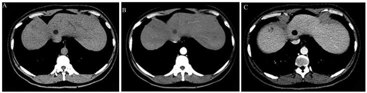
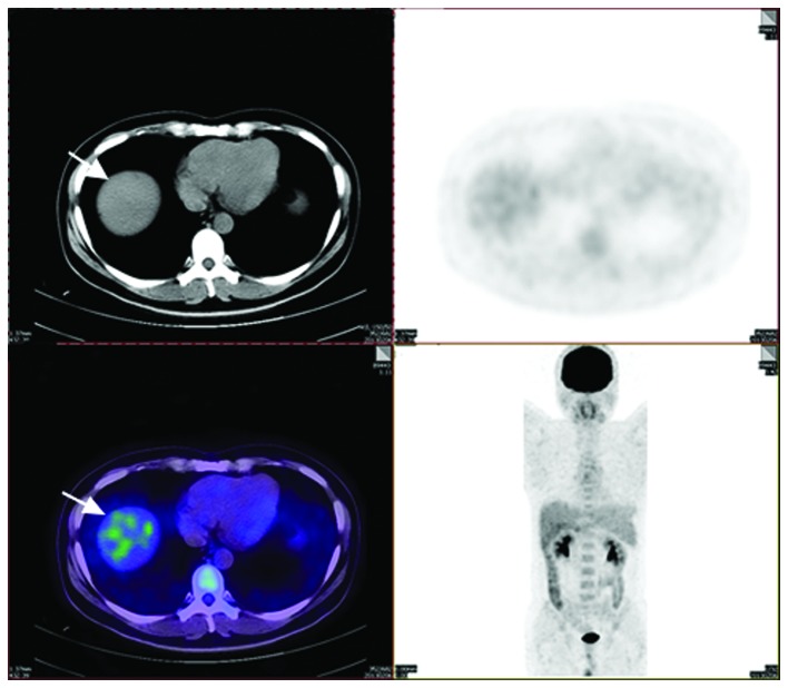
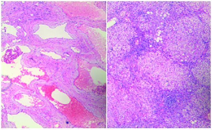
A 47-year-old male presented with a six-month history of fatigue and a four-month history of alanine and aspartate aminopherase elevation. Laboratory examination revealed that the serum α-fetoprotein (AFP) level was 371.51 μg/l (normal range, 0-20 μg/l), and a computed tomography scan revealed a hypodense lesion in the left hepatic lobe. During laparotomy, a dark red-colored soft tumor (1.5×1.7 cm in diameter) was found in segment eight of the liver. Intra-operative pathology and post-operative histopathology examinations revealed that the tumor was a hepatic cavernous hemangioma. The serum AFP level was decreased to 24.45 μg/l by the second post-operative week. The literature was searched and only three similar cases were found. A brief review of this rare disease entity was produced, which attempted to explain this appearance reasonably.
Keywords: cancer stem cell; hepatic cavernous hemangioma; α-fetoprotein.
Figures
References
-
- Ishak KG, Anthony PP, Niederau C, Nakanuma Y. Mesenchymal tumours of the liver. In: Hamilton SR, Aaltonen LA, editors. World Health Organization Classification of Tumors. Pathology and Genetics of Tumours of the Digestive System. IARC Press; Lyon, France: 2000. pp. 191–198.
LinkOut - more resources
Full Text Sources
Other Literature Sources