

Metastasizing leiomyoma to heart
- PMID: 25624981
- PMCID: PMC4300065
- DOI: 10.14797/mdcj-10-4-251
Metastasizing leiomyoma to heart
Abstract
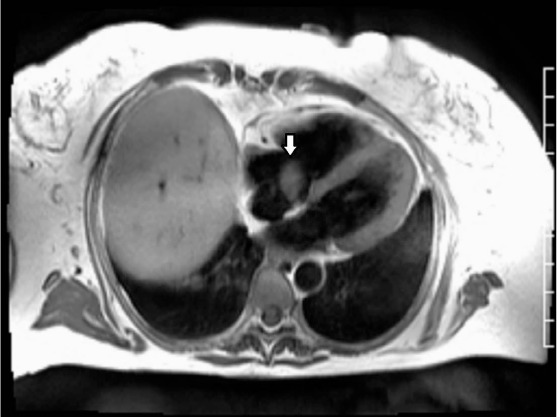
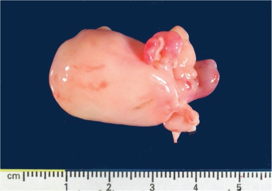
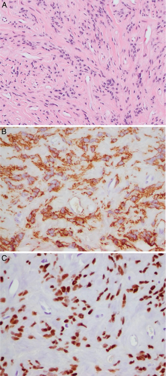
Cardiac smooth muscle tumors are rare. Three different clinical settings for these tumors have been reported, including benign metastasizing leiomyoma from the uterus, primary cardiac leiomyoma and leiomyosarcoma, and intravenous cardiac extension of pelvic leiomyoma, which is the most common. We present a case of a 55-year-old woman with a benign metastasizing leiomyoma to the heart 17 years after hysterectomy and 16 years after metastasis to the lung. Immunohistochemical stains for smooth muscle actin, desmin, and estrogen and progesterone receptors were positive, indicating a smooth muscle tumor of uterine origin. To our knowledge, this is only the fourth reported case of benign metastasizing leiomyoma to the heart and the first case of long-delayed cardiac metastasis after successful treatment of pulmonary metastasis. It illustrates that benign metastasizing leiomyoma should be included in the differential diagnosis of cardiac tumors in patients with a history of uterine leiomyoma, especially when associated with pulmonary metastasis.
Keywords: benign metastasizing leiomyoma; heart; primary cardiac tumor.
Figures

References
-
- Galvin SD, Wademan B, Chu J, Bunton RW. Benign metastasizing leiomyoma: a rare metastatic lesion in the right ventricle. Ann Thorac Surg. 2010 Jan;89(1):279–81. - PubMed
-
- Kocica MJ, Vranes MR, Kostic D, Kovacevic-Kostic N, Lackovic V, Bozic-Mihajlovic V et al. Intravenous leiomyomatosis with extension to the heart: rare or underestimated? J Thorac Cardiovasc Surg. 2005 Dec;130(6):1724–6. - PubMed
-
- Takemura G, Takatsu Y, Kaitani K, Ono M, Ando F, Tanada S et al. Metastasizing uterine leiomyoma. A case with cardiac and pulmonary metastasis. Pathol Res Pract. 1996 Jun;192(6):622–9. discussion 630–3. - PubMed
-
- Thukkani N, Ravichandran PS, Das A, Slater MS. Leiomyomatosis metastatic to the tricuspid valve complicated by pelvic hemorrhage. Ann Thorac Surg. 2005 Feb;79(2):707–9. - PubMed
-
- Qin C, Chen L, Xiao YB, Chen BC. Giant primary leiomyoma of the right ventricle. J Card Surg. 2010 Mar;25(2):169–171. - PubMed
Publication types
MeSH terms
LinkOut - more resources
Full Text Sources
Other Literature Sources
Medical
