

α-Tomatine inhibits growth and induces apoptosis in HL-60 human myeloid leukemia cells
- PMID: 25625536
- PMCID: PMC4735690
- DOI: 10.3892/mmr.2015.3238
α-Tomatine inhibits growth and induces apoptosis in HL-60 human myeloid leukemia cells
Abstract
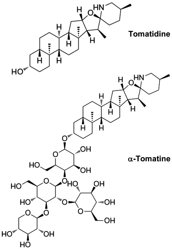
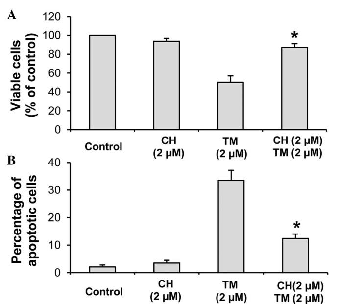
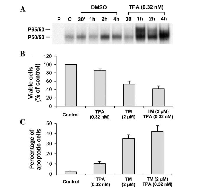
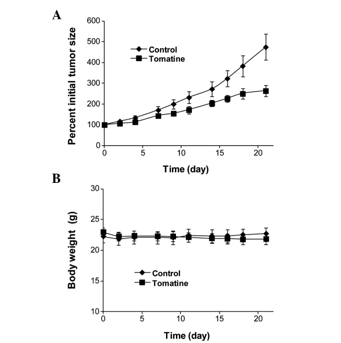
α‑Tomatine is a glycoalkaloid that occurs naturally in tomatoes (Lycopersicon esculentum). In the present study, the effects of α‑tomatine on human myeloid leukemia HL‑60 cells were investigated. Treatment of HL‑60 cells with α‑tomatine resulted in growth inhibition and apoptosis in a concentration‑dependent manner. Tomatidine, the aglycone of tomatine had little effect on the growth and apoptosis of HL‑60 cells. Growth inhibition and apoptosis induced by α‑tomatine in HL‑60 cells was partially abrogated by addition of cholesterol indicating that interactions between α‑tomatine and cell membrane‑associated cholesterol may be important in mediating the effect of α‑tomatine. Activation of nuclear factor‑κB by the phorbol ester, 12‑O‑tetradecanoylphorbol‑13‑acetate failed to prevent apoptosis in HL‑60 cells treated with α‑tomatine. In animal experiments, it was found that treatment of mice with α‑tomatine inhibited the growth of HL‑60 xenografts in vivo. Results from the present study indicated that α‑tomatine may have useful anti‑leukemia activities.
Figures


Similar articles
-
Combination of α-Tomatine and Curcumin Inhibits Growth and Induces Apoptosis in Human Prostate Cancer Cells.PLoS One. 2015 Dec 2;10(12):e0144293. doi: 10.1371/journal.pone.0144293. eCollection 2015. PLoS One. 2015. PMID: 26630272 Free PMC article.
-
α-Tomatine-mediated anti-cancer activity in vitro and in vivo through cell cycle- and caspase-independent pathways.PLoS One. 2012;7(9):e44093. doi: 10.1371/journal.pone.0044093. Epub 2012 Sep 6. PLoS One. 2012. PMID: 22970166 Free PMC article.
-
Enhancement of TPA-induced growth inhibition and apoptosis in myeloid leukemia cells by BAY 11-7082, an NF-kappaB inhibitor.Int J Oncol. 2005 Oct;27(4):941-8. Int J Oncol. 2005. PMID: 16142309
-
The steroidal alkaloids α-tomatine and tomatidine: Panorama of their mode of action and pharmacological properties.Steroids. 2021 Dec;176:108933. doi: 10.1016/j.steroids.2021.108933. Epub 2021 Oct 23. Steroids. 2021. PMID: 34695457 Review.
-
Physiological functions, pharmacological aspects and nutritional importance of green tomato- a future food.Crit Rev Food Sci Nutr. 2024;64(27):9711-9739. doi: 10.1080/10408398.2023.2212766. Epub 2023 Jun 2. Crit Rev Food Sci Nutr. 2024. PMID: 37267154 Review.
Cited by
-
Agri-Food Waste Recycling for Healthy Remedies: Biomedical Potential of Nutraceuticals from Unripe Tomatoes (Solanum lycopersicum L.).Foods. 2024 Jan 20;13(2):331. doi: 10.3390/foods13020331. Foods. 2024. PMID: 38275698 Free PMC article. Review.
-
The metabolic changes that effect fruit quality during tomato fruit ripening.Mol Hortic. 2022 Jan 20;2(1):2. doi: 10.1186/s43897-022-00024-1. Mol Hortic. 2022. PMID: 37789428 Free PMC article. Review.
-
Therapeutic Potential of Solanum Alkaloids with Special Emphasis on Cancer: A Comprehensive Review.Drug Des Devel Ther. 2024 Jul 16;18:3063-3074. doi: 10.2147/DDDT.S470925. eCollection 2024. Drug Des Devel Ther. 2024. PMID: 39050799 Free PMC article. Review.
-
Main chemical constituents and mechanism of anti-tumor action of Solanum nigrum L.Cancer Med. 2024 Aug;13(16):e7314. doi: 10.1002/cam4.7314. Cancer Med. 2024. PMID: 39155844 Free PMC article. Review.
-
The role of cholesterol metabolism in leukemia.Blood Sci. 2019 Sep 17;1(1):44-49. doi: 10.1097/BS9.0000000000000016. eCollection 2019 Aug. Blood Sci. 2019. PMID: 35402792 Free PMC article. Review.
References
Publication types
MeSH terms
Substances
Grants and funding
LinkOut - more resources
Full Text Sources
Other Literature Sources
