Review
. 2015 Jan 23;5(1):294-320.
doi: 10.3390/life5010294.

What RNA World? Why a Peptide/RNA Partnership Merits Renewed Experimental Attention

Affiliations
Review

What RNA World? Why a Peptide/RNA Partnership Merits Renewed Experimental Attention

Charles W Carter. Life (Basel). .

Abstract

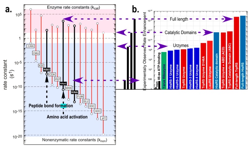
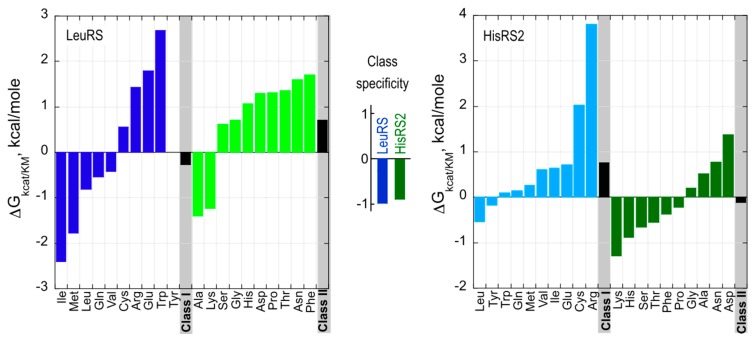
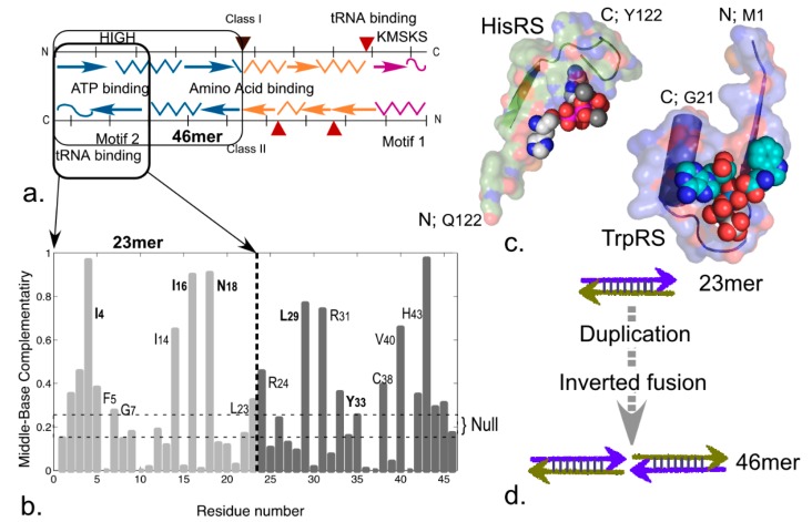
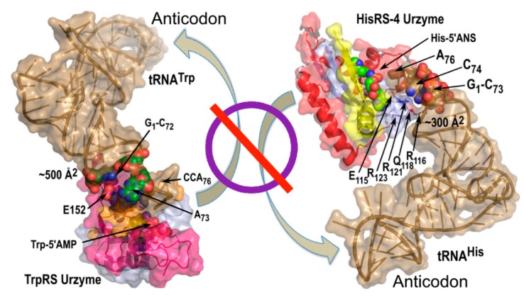
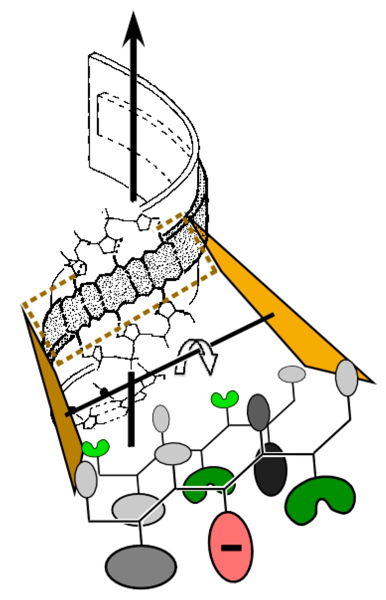
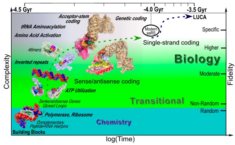
We review arguments that biology emerged from a reciprocal partnership in which small ancestral oligopeptides and oligonucleotides initially both contributed rudimentary information coding and catalytic rate accelerations, and that the superior information-bearing qualities of RNA and the superior catalytic potential of proteins emerged from such complexes only with the gradual invention of the genetic code. A coherent structural basis for that scenario was articulated nearly a decade before the demonstration of catalytic RNA. Parallel hierarchical catalytic repertoires for increasingly highly conserved sequences from the two synthetase classes now increase the likelihood that they arose as translation products from opposite strands of a single gene. Sense/antisense coding affords a new bioinformatic metric for phylogenetic relationships much more distant than can be reconstructed from multiple sequence alignments of a single superfamily. Evidence for distinct coding properties in tRNA acceptor stems and anticodons, and experimental demonstration that the two synthetase family ATP binding sites can indeed be coded by opposite strands of the same gene supplement these biochemical and bioinformatic data, establishing a solid basis for key intermediates on a path from simple, stereochemically coded, reciprocally catalytic peptide/RNA complexes through the earliest peptide catalysts to contemporary aminoacyl-tRNA synthetases. That scenario documents a path to increasing complexity that obviates the need for a single polymer to act both catalytically and as an informational molecule.


Figures

Figure 1
Figure 1
Stereochemistry of peptide-RNA conformational complementarity [1]. The minor groove in double-stranded RNA (magenta) complements the preferred right-hand twist of antiparallel β hairpin structures (wheat). Adjacent nucleotide and peptide strands are parallel (5'-3'; N-C) and the two sets are antiparallel. Van der Waals distances between peptide and nucleotide components are optimal precisely at a peptide radius for which there are exactly two amino acids per base. (Inset) The double-double helix is also stabilized by recurring hydrogen bonds between peptide carbonyl and the ribose 2'OH groups and between amide nitrogens and water molecules (blue spheres) between the ribose O1 and 2'OH groups. The resulting hydrogen-bonded network stabilizes a ribose orientation such that the 3'OH group is poised to serve as a nucleophile for polymerization.
Figure 2
Figure 2
Templating and reciprocal autocatalysis proposed by Carter and Kraut. Rudimentary coding and catalytic surfaces are evident in both oligomers. Screw dislocations of peptide strands relative to nucleotide strands create pockets for the addition of new units—either a dipeptide or a nucleotide. (a) Addition of amino acid or dipeptide. RNA strands afford a scaffold allowing one peptide strand (teal) to serve as template and the other (wheat) as primer; (b) Addition of a nucleotide. Antiparallel β-structure affords a scaffold on which one RNA strand (yellow) functions as template while the other (magenta) functions as primer.
Figure 3
Figure 3
Urzymes isolated from Class I TrpRS (130 amino acids) and Class II HisRS (124 amino acids). (a) Monomer architectures. Both enzymes are dimeric. Monomers consist of two consensus domains, catalytic and anticodon-binding (ABD). The 46-amino acid ATP binding sites are blue and bounded by transparent surfaces; the remainder of the Urzymes are red. Catalytic domains of both also include insertions, colored amber. Active-site ligands are shown as spheres; (b) Secondary structures are dissimilar; Class I is a Rossmann fold with parallel β-strands; Class II is an antiparallel structure; (c) Amino acid (yellow) and ATP (cyan) substrates are spheres. Stick models of Class I-defining signatures PxxxxHIGH (green) and KMSKS (red) and Class II Motif 2 (green) are surrounded by transparent surfaces to reveal catalytically important interactions with ATP. The cartoons are based on crystal structures of the full-length enzymes. However, long-time MD simulations of both Urzymes in the presence of both substrates have shown that the structures shown here persist, but that in the absence of tryptophan the Class I specificity helix above the bound tryptophan re-orients, removing several key interactions involved in specific recognition [21,25,26].
Figure 4
Figure 4
Relationship between TrpRS Urzyme and connecting peptide 1 (CP1). The alpha carbons to which the N and C termini of the insertion attach are separated by almost exactly the distance of a peptide bond (blue dots). This enables its removal by protein engineering, which was accomplished using the Rosetta design program [15,21]. The dashed arrow indicates where the anticodon-binding domain attaches (Adapted from [15]).
Figure 5
Figure 5
Quantitative framework in which to assess the catalytic significance of Urzymes and various other putative stages of aaRS evolution. (a) Rate accelerations estimated from experimental data for single substrate (red) and bi-substrate (Black, Bold) reactions adapted from [31] to include uncatalyzed and catalyzed rates of bi-substrate reactions of the ribosome [22,23], amino acid activation [32], and kinases [33]. Second-order rate constants (black bars) were converted into comparable units by multiplying by 0.002 M, which is the ATP concentration used to assay the catalysts shown in B; (b) Experimental rate accelerations for amino acid activation, estimated from steady-state kinetics of 32PPi exchange as kcat/KM for catalysts derived from Class I and Class II aminoacyl-tRNA synthetases [14,21]. Vertical scales in (a) and (b) are the same, and the origin of the histogram in (b) is set equal to the uncatalyzed rate of amino acid activation (AAact) in (a). Red bars denote Class I Tryptophanyl- and Leucyl-tRNA synthetase constructs, blue bars denote Class II Histidyl-tRNA synthetase constructs, black vertical lines denote catalysis by 46-residue ATP binding sites, and green denotes a ribozymal catalyst [34] for a different amino acid activation reaction, included for comparison. Research presented in (a), (b) was originally published in [13]. © The American Society for Biochemistry and Molecular Biology.
Figure 6
Figure 6
Amino acid specificity spectra of Class I LeuRS and Class II HisRS2 Urzymes. The net free energy for specificity of Class I and II Urzymes for homologous vs. heterologous substrates, i.e., ΔG(kcat/KM)ref – ΔG(kcat/KM)amino acid (i), where ref is the cognate amino acid, is approximately 1 kcal/mole for both Class I and II Urzymes (center). Class I amino acids are colored blue; Class II amino acids are colored green. Bold colors denote substrates from the homologous Class; pastel colors denote heterologous substrates.
Figure 7
Figure 7
Evidence for an inverted repeat in coding for Class I ATP binding sites. (a) Schematic of functional divisions in the Class I and II Urzymes, showing parallel localization of ATP, amino acid, and tRNA binding sites; (b) Coding sequences for the 46 residue segment have significantly elevated middle-base pairing (0.28 ± 0.0005 vs. 0.25 ± 0.0004) when aligned antisense to each other. Frequencies of middle-base pairing between residues 4 and 43 together with 16, 18 and 29, 31 and other pairwise comparisons along the gene are evidence of coding by an ancestral palindrome; (c) Structures corresponding to the sense/antisense 23mer containing Class I PxxxHIGH and Class II Motif 2 binding determinants; (d) Putative evolution of the 46mer gene via formation of an inverted repeat. Such a gene would have been stable either as a duplex or as an RNA hairpin.
Figure 8
Figure 8
Urzyme size precludes tRNA anticodon recognition. Urzyme interactions include binding determinants for the tRNA acceptor stem, but cannot interact with the anticodon.
Figure 9
Figure 9
Possible relevance of mass, β-branching, and carboxylates to the operational RNA code (adapted from [56]). For ancient β-hairpins to interact with double-stranded RNA as envisioned by Carter and Kraut [1], large side chains would necessarily have faced away from the RNA minor groove. β-branched side chains on either face (re-entrant angles; green; threonine, valine on the inward face; isoleucine on the outer face) enhance β-structure formation. Carboxylate side chains in outward facing positions (red) could enhance solubility [58,59] and coordinate catalytic divalent metals, either for catalysis or to protect against RNA degradation [60].
Figure 10
Figure 10
Detailed scenario for the emergence of peptide and RNA polymers and steps toward the creation of the genetic code. Approximate time intervals run from the formation of liquid water on the earth to the appearance of the last universal common ancestor (LUCA) and specifically plot stages in the evolution of the aaRS that we have documented either by model building (Figure 1 and Figure 2) or experiments (Figure 5), and which therefore are arguable intermediates that do not require prior evolution of human technology. Major biochemical events are in italicized bold face. Sense/antisense coding appeared with the first reciprocally autocatalytic peptide–RNA complex and persisted until the evolution of aminoacyl-tRNA synthetases, tRNAs, and ribosomes enabled higher-specificity genetic coding.

References

    1. Carter C.W., Jr., Kraut J. A Proposed Model for Interaction of Polypeptides with RNA. Proc. Natl. Acad. Sci. USA. 1974;71:283–287. doi: 10.1073/pnas.71.2.283. - DOI - PMC - PubMed
    1. Carter C.W., Jr. Cradles for Molecular Evolution. New Scientist. 1975 Mar 27;:784–787.
    1. Joyce G., Orgel L.E. Progress Toward Understanding the Origin of the RNA World. In: Gesteland R.F., Cech T.R., Atkins J., editors. The RNA World. 3rd ed. Cold Spring Harbor Laboratory; Cold Spring Harbor, NY, USA: 2006.
    1. Akst J. RNA World 2.0. The Scientist. 2014 Mar 1;:34–40.
    1. Watson J.D., Crick F.H.C. A Structure for Deoxyribose Nucleic Acid. Nature. 1953;171:737–738. doi: 10.1038/171737a0. - DOI - PubMed

LinkOut - more resources