. 2015 Jun 15;32(12):893-907.
doi: 10.1089/neu.2014.3718. Epub 2015 May 5.

Spinal interneurons and forelimb plasticity after incomplete cervical spinal cord injury in adult rats

Affiliations

Spinal interneurons and forelimb plasticity after incomplete cervical spinal cord injury in adult rats

Elisa Janine Gonzalez-Rothi et al. J Neurotrauma. .

Abstract

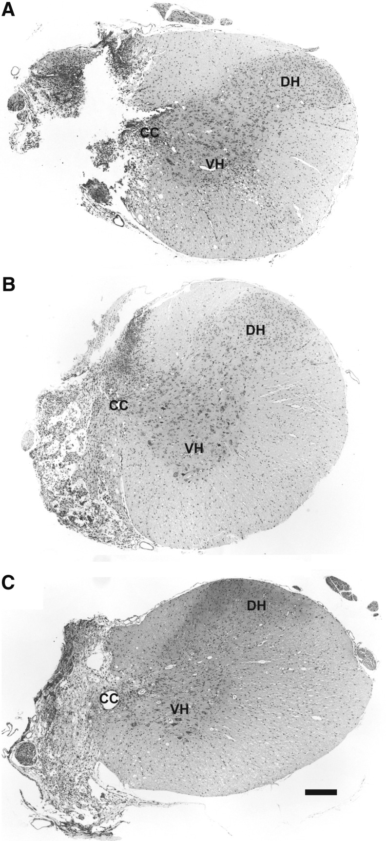
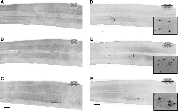
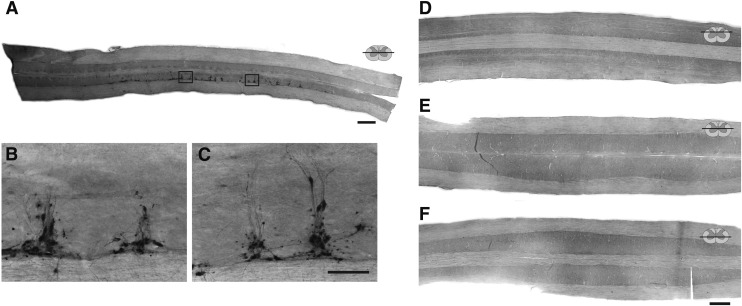
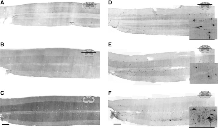
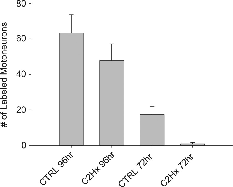
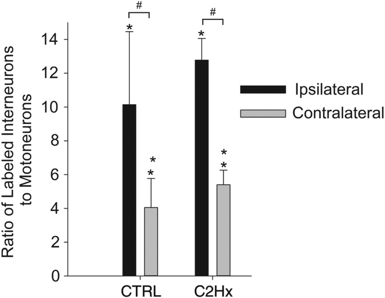
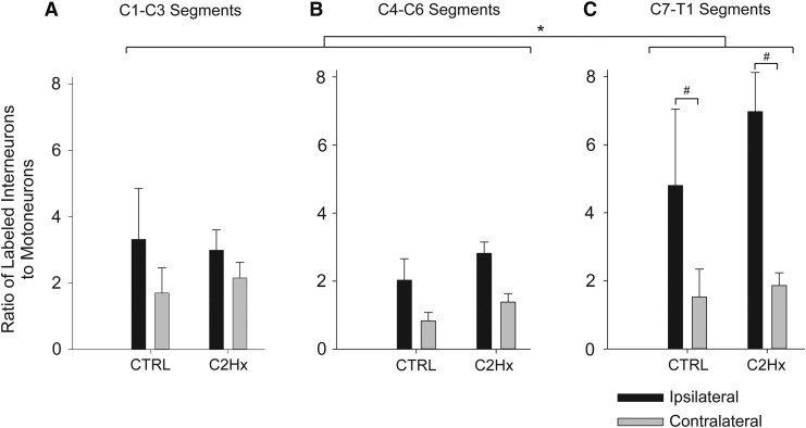
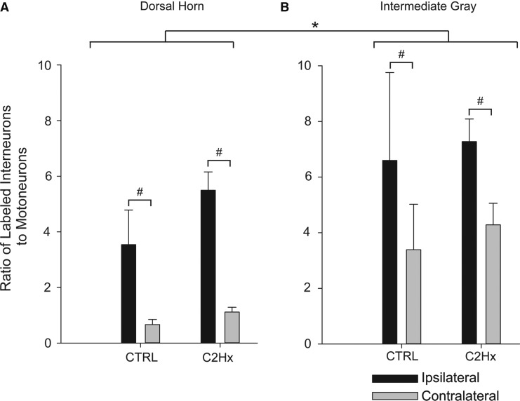
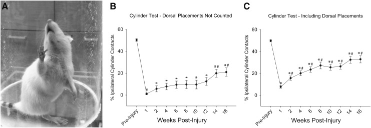
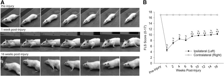
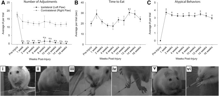
Cervical spinal cord injury (cSCI) disrupts bulbospinal projections to motoneurons controlling the upper limbs, resulting in significant functional impairments. Ongoing clinical and experimental research has revealed several lines of evidence for functional neuroplasticity and recovery of upper extremity function after SCI. The underlying neural substrates, however, have not been thoroughly characterized. The goals of the present study were to map the intraspinal motor circuitry associated with a defined upper extremity muscle, and evaluate chronic changes in the distribution of this circuit following incomplete cSCI. Injured animals received a high cervical (C2) lateral hemisection (Hx), which compromises supraspinal input to ipsilateral spinal motoneurons controlling the upper extremities (forelimb) in the adult rat. A battery of behavioral tests was used to characterize the time course and extent of forelimb motor recovery over a 16 week period post-injury. A retrograde transneuronal tracer - pseudorabies virus - was used to define the motor and pre-motor circuitry controlling the extensor carpi radialis longus (ECRL) muscle in spinal intact and injured animals. In the spinal intact rat, labeling was observed unilaterally within the ECRL motoneuron pool and within spinal interneurons bilaterally distributed within the dorsal horn and intermediate gray matter. No changes in labeling were observed 16 weeks post-injury, despite a moderate degree of recovery of forelimb motor function. These results suggest that recovery of the forelimb function assessed following C2Hx injury does not involve recruitment of new interneurons into the ipsilateral ECRL motor pathway. However, the functional significance of these existing interneurons to motor recovery requires further exploration.

Keywords: SCI; functional plasticity; propriospinal interneurons; upper extremity.


Figures

<b>FIG. 1.</b>
FIG. 1.
Representative histological sections illustrating verified complete high cervical lateral hemisection (C2Hx) lesions 4 μm transverse sections at C2 taken from rats, 1 (A), 2 (B), and 8 weeks post-injury (C) stained with cresyl violet. The absence of healthy white and gray matter in the ipsilateral spinal cord suggests anatomically complete C2Hx lesions. CC, central canal; DH, dorsal horn; VH, ventral horn. Scale Bar: 200 μm.
<b>FIG. 2.</b>
FIG. 2.
Representative longitudinal (horizontal) sections through the cervical spinal cord of uninjured adult female Sprague–Dawley rats, 72 (A–C) and 96 (D–F) h following injection of pseudorabies virus (PRV) into the left extensor carpi radialis longus (ECRL) muscle. Sections have been immunolabeled for the presence of PRV. Low resolution images (A–F) demonstrate the distribution of PRV labeling in the cervical spinal cord, and high resolution images (insets, panels D–F) demonstrate ECRL motoneuron and interneuron morphology. ECRL motoneuron labeling in the ventral horn (C and F), as well as interneuronal labeling in the intermediate gray matter (B and E) and the dorsal horn (A and D) of the cervical spinal cord at the 96 h post-injection time point. Rostrocaudal orientation is from left to right. Scale bars: 1 mm (panels A–F) and 100 μm (inset panels).
<b>FIG. 3.</b>
FIG. 3.
Representative longitudinal (horizontal) sections through the thoracic (A–C) and cervical spinal (D–F) cord of uninjured adult female Sprague–Dawley rats, 72 h after injection of pseudorabies virus (PRV) into the left extensor carpi radialis longus (ECRL) muscle. These control experiments were conducted to determine whether the PRV-positive labeling observed in the cervical spinal cord after injection into the ECRL muscle (see Fig. 2) was associated with non-ECRL circuitry (i.e., sympathetic labeling). In these control experiments, the left radial nerve was cut prior to injection of PRV, preventing retrograde labeling via ECRL motoneurons. Sections have been immunolabeled for the presence of PRV. Sections A–C demonstrate the distribution of sympathetic pre-ganglionic (non-ECRL) labeling associated with PRV injection into the ECRL muscle, which was concentrated within in the intermediolateral gray matter of the thoracic spinal cord. Sections D–F demonstrate the absence of PRV labeling in the dorsal horn (D), the intermediate gray matter (E), or the ventral horn (F) of the cervical spinal cord following radial nerve section, confirming that cervical labeling observed in the PRV-tracing experiments is associated with the ECRL circuitry. Rostrocaudal orientation is from left to right. Scale bars: 1 mm (A, D–F) and 100 μm (B and C).
<b>FIG. 4.</b>
FIG. 4.
Representative longitudinal (horizontal) sections through the cervical spinal cord of rats, 16 weeks after high cervical lateral hemisection (C2Hx) injury, 72 (A–C) and 96 (D–F) h after injection of pseudorabies virus (PRV) into the left extensor carpi radialis longus (ECRL) muscle. Sections have been immunolabeled for the presence of PRV. Low resolution images (A–F) demonstrate the distribution of PRV labeling in the cervical spinal cord, and high resolution images (insets, panels D–F) demonstrate ECRL motoneuron and interneuron morphology. Motoneuron labeling was evident in the ipsilateral ventral horn (C and F), as well as bilateral interneuronal labeling in the intermediate gray matter (B and E) and dorsal horns (A and D) at the 96 h post-injection time point. As compared with uninjured controls, first-order PRV labeling at the 72 h time point was reduced, although at 96 h, significant interneuronal labeling was evident, extending throughout the cervical spinal cord in both the dorsal horn and the intermediate gray matter (D–E). Rostrocaudal orientation is from left to right. Scale bars: 1 mm (panels A–F) and 100 μm (inset panels).
<b>FIG. 5.</b>
FIG. 5.
The number of pseudorabies virus (PRV)-positive extensor carpi radialis longus (ECRL) motoneurons (± standard error of the mean [SEM]) in the cervical spinal cord of control (CTRL) and high cervical lateral hemisection (C2Hx) rats. The number of labeled motoneurons in both CTRL and C2Hx groups was significantly greater (p<0.05) 96 h after delivery of PRV to the left ECRL than at 72 h (*). No evidence of contralateral motoneuron labeling was observed at any time point, in any group.
<b>FIG. 6.</b>
FIG. 6.
Pre-motor extensor carpi radialis longus (ECRL) interneurons in the cervical spinal cord. The number of PRV-positive cells counted are expressed relative to the number of PRV-labeled motoneurons (± standard error of the mean [SEM]). The number of labeled interneurons in both control (CTRL) and high cervical lateral hemisection (C2Hx) groups was significantly greater (p<0.05) 96 h after delivery of pseudorabies virus (PRV) to the left ECRL than at 72 h (*); 72 h after delivery of PRV, the number of ipsilateral labeled cells was no different than the number of contralateral cells in both CTRL and C2Hx groups; 96 h after delivery of PRV, the number of ipsilateral labeled cells was significantly greater (p<0.001) than the number of contralateral cells in both CTRL and C2Hx groups (#). There was no difference in the number of labeled cells between CTRL and C2Hx groups at either time point.
<b>FIG. 7.</b>
FIG. 7.
The rostrocaudal distribution of pre-motor extensor carpi radialis longus (ECRL) Interneurons in the cervical spinal cord. The number of pseudorabies (PRV)-positive cells counted in each segment are expressed relative to the number of PRV-labeled motoneurons (± standard error of the mean [SEM]). The number of labeled interneurons in both control (CTRL) and high cervical lateral hemisection (C2Hx) groups was significantly greater (p<0.05) 96 hours after delivery of PRV to the left ECRL than at 72 h at all levels (*); 72 h after delivery of PRV, the number of ipsilateral labeled cells was no different than the number of contralateral cells at any level in both CTRL and C2Hx groups; 96 h after delivery of PRV, the number of ipsilateral labeled cells was significantly greater (p<0.001) than the number of contralateral cells at C7-T1 in both CTRL and C2Hx groups (#). There was no difference in the number of labeled cells between CTRL and C2Hx groups at any level, at either time point.
<b>FIG. 8.</b>
FIG. 8.
The regional distribution of pre-motor extensor carpi radialis longus (ECRL) interneurons in the cervical spinal cord. The number of pseudorabies virus (PRV)-positive cells counted in the dorsal horn (A) and in intermediate gray matter (B) are expressed relative to the number of PRV-labeled motoneurons (± standard error of the mean [SEM]). The number of labeled interneurons in both control (CTRL) and high cervical lateral hemisection (C2Hx) groups was significantly greater (p<0.05) 96 h after delivery of PRV to the left ECRL than at 72 hin both the dorsal horn and in the intermediate gray matter (*); 72 h after delivery of PRV, the number of ipsilateral labeled cells was no different than the number of contralateral cells in either region in both CTRL and C2Hx groups; 96 h after delivery of PRV, the number of ipsilateral labeled cells was significantly greater (p<0.001) than the number of contralateral cells in both the CTRL and the C2Hx groups (#). There was no difference in the number of labeled cells between CTRL and C2Hx groups in either region, at either time point.
<b>FIG. 9.</b>
FIG. 9.
(A) The impact of high cervical lateral hemisection (C2Hx) on ipsilateral upper extremity use during vertical cylinder exploration. Ipsilateral paw use was represented as the percentage of ipsilateral placements relative to the total number of placements. Scores were calculated both with (Panel C) and without (Panel B) dorsal paw placements included in the quantification. All post-injury data points were significantly different than pre-injury values (*p<0.05). If dorsal paw placements were excluded from the quantification, progressive improvements in ipsilateral forelimb use occurred beginning in week 14 (#p<0.05). If dorsal paw placements were included in the quantification, progressive improvements in ipsilateral forelimb use occurred beginning in week 2 (#p<0.05). Values are mean ± standard error (SE) using one way repeated measures analysis of variance (RM ANOVA). p<0.05.
<b>FIG. 10</b>
FIG. 10
(A) The impact of high cervical lateral hemisection (C2Hx) on ipsilateral locomotor function was assessed during open field-locomotion using the Forelimb Locomotor Scale (FLS). Representative images depict locomotor function prior to and at 1 and 16 weeks post-injury. (B) FLS scores were determined in uninjured rats and at 1, 2, 4, 6, 8, 10, 12, 14, and 16 weeks post-C2Hx injury. All post-injury ipsilateral data points were significantly different than the contralateral (%p<0.05) scores, and were significantly different than pre-injury values (*p<0.05). Progressive improvements in forelimb locomotor function occurred beginning in week 8, with each successive data point increasing significantly from the 6 week time point (#p<0.05). Values are mean ± SE using one-way repeated measures analysis of variance (RM ANOVA). p<0.05.
<b>FIG. 11.</b>
FIG. 11.
Vermicelli pasta handling test. The average number of paw adjustments (A), time to eat (B), and number of atypical behaviors (C and D) were quantified (mean ± standard error of the mean [SEM]). (A) The average number of adjustments made by the ipsilateral (left) paw was dramatically reduced after injury (p<0.05) and was significantly lower than the number of adjustments made by the contralateral (right) paw (p<0.05). Rats did not regain ipsilateral paw use over the course of 16 weeks (p<0.05). (B) No difference in the time to eat the pasta was observed, although there was considerable variability between animals and between testing sessions. (C) The average number of atypical behaviors observed per trial increased following high cervical lateral hemisection (C2Hx) injury (p<0.05). %significantly different from contralateral; *significantly different from pre-injury; &significantly different from 4 weeks post-C2Hx; $significantly different from 6 weeks post-C2Hx. (D) Examples of normal (i) and the most common atypical behaviors (ii-vi) demonstrated by rats during the vermicelli pasta handling test. Example images include normal eating behavior (i), head tilt with angled pasta hold (ii, iii, iv and vi), hunched posture (iii, iv, and v), and failure to contact (ii, iii, iv, and vi). Atypical behaviors were observed frequently after injury, and may represent compensatory strategies for accomplishing the pasta eating task.

Similar articles

Cited by

References

    1. Lane M.A., White T.E., Coutts M.A., et al. (2008). Cervical prephrenic interneurons in the normal and lesioned spinal cord of the adult rat. J. Comp. Neurol. 511, 692–709 - PMC - PubMed
    1. Reier P., Lane M., Hall E.D., Teng Y.D., and Howland D.R. (2012). Translational spinal cord injury research: preclinical guidelines and challenges. Handb. Clin. Neurol. 109, 411–433 - PMC - PubMed
    1. Vizzard M., Erickson V., Card J., Roppolo J., and de Groat W. (1995). Transneuronal labeling of neurons in the adult rat brainstem and spinal cord after injection of pseudorabies virus into the urethra. J. Comp. Neurol. 355, 629–640 - PubMed
    1. Hou S., Duale H., Cameron A.A., Abshire S.M., Lyttle T.S., and Rabchevsky A.G. (2008). Plasticity of lumbosacral propriospinal neurons is associated with the development of autonomic dysreflexia after thoracic spinal cord transection. J. Comp. Neurol. 509, 382–399 - PMC - PubMed
    1. Billig I., Forris J.M., Enquist L.W., Card J., and Yates B.J. (2000). Definition of neuronal circuitry controlling the activity of phrenic and abdominal motoneurons in the ferret using recombinant strains of pseudorabies virus. J. Neurosci. 20, 7446–7454 - PMC - PubMed

Publication types

LinkOut - more resources