Review
. 2014;11(12):1597-607.
doi: 10.1080/15476286.2015.1004955.

RNA methyltransferases involved in 5' cap biosynthesis

Affiliations
Review

RNA methyltransferases involved in 5' cap biosynthesis

Magdalena Byszewska et al. RNA Biol. 2014.

Abstract

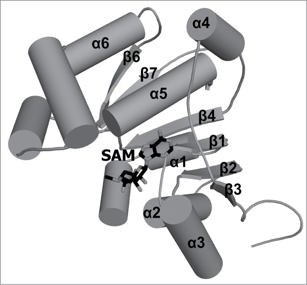
In eukaryotes and viruses that infect them, the 5' end of mRNA molecules, and also many other functionally important RNAs, are modified to form a so-called cap structure that is important for interactions of these RNAs with many nuclear and cytoplasmic proteins. The RNA cap has multiple roles in gene expression, including enhancement of RNA stability, splicing, nucleocytoplasmic transport, and translation initiation. Apart from guanosine addition to the 5' end in the most typical cap structure common to transcripts produced by RNA polymerase II (in particular mRNA), essentially all cap modifications are due to methylation. The complexity of the cap structure and its formation can range from just a single methylation of the unprocessed 5' end of the primary transcript, as in mammalian U6 and 7SK, mouse B2, and plant U3 RNAs, to an elaborate m(7)Gpppm(6,6)AmpAmpCmpm(3)Um structure at the 5' end of processed RNA in trypanosomes, which are formed by as many as 8 methylation reactions. While all enzymes responsible for methylation of the cap structure characterized to date were found to belong to the same evolutionarily related and structurally similar Rossmann Fold Methyltransferase superfamily, that uses the same methyl group donor, S-adenosylmethionine; the enzymes also exhibit interesting differences that are responsible for their distinct functions. This review focuses on the evolutionary classification of enzymes responsible for cap methylation in RNA, with a focus on the sequence relationships and structural similarities and dissimilarities that provide the basis for understanding the mechanism of biosynthesis of different caps in cellular and viral RNAs. Particular attention is paid to the similarities and differences between methyltransferases from human cells and from human pathogens that may be helpful in the development of antiviral and antiparasitic drugs.

Keywords: RNA maturation; RNA modification; antiviral drugs; cap; crystallography; mRNA; methylation; modified nucleotides; post-transcriptional modification; trypanosomes.


Figures

Figure 1.
Figure 1.
Schematic representation of the conserved core of Rossmann-fold Methyltransferase (RFM) catalytic domains. The β-sheet is composed of 7 β-strands (gray arrows) surrounded by 6 α-helices (semi-transparent tubes) forms the fold that is typical for SAM-dependent methyltransferases. All secondary structure elements of the conserved core are labeled as α1, β1, etc. The stars indicate points of most frequent insertions and terminal fusions with other domains.
Figure 2.
Figure 2.
Crystal structure of the cap0 methyltransferase from Encephalitozoon cuniculi. A stick representation of the ligands bound to cap0 methyltransferase. The guanosine cap analog position was defined based on the structure deposited as 1RI2 in the PDB, and the methyl group donor position was depicted based on the structure deposited as 1RI4 in the PDB. Secondary structure elements that correspond to elements of the conserved RFM core are labeled. Secondary structure elements outside of the conserved core are not labeled.
Figure 3.
Figure 3.
Comparison of the crystal structures of 2-O-ribose methyltransferases. (A) Superimposition of the catalytic domain of human CMTr1 methyltransferase (colored black; PDB ID: 4N48), VP39 methyltransferase from the vaccinia virus (colored dark gray; PDB ID: 1AV6) and the NS5 protein from the Wesselsbron virus (colored bright gray; PDB ID: 3EMB). The ligands are shown in stick representation and they are colored corresponding to the hue used for protein molecules representation. Secondary structure elements that correspond to elements of the conserved RFM core are labeled (α1, β6, and β7 are hidden behind other elements and their labels have been omitted). Secondary structure elements outside of the conserved core are not labeled. (B) The capped oligoribonucleotide (m7GpppGAUC) located in its binding pocket on the surface of human CMTr1 MTase is shown in stick representation. The side chains of Phe206 and Glu373 that correspond to stacking residues in viral methyltransferases and the 3 catalytic residues are also displayed. (C) The crystal structure of the VP39 methyltransferase from vaccinia virus in complex with m7GpppGAAAAA (shown in stick representation). The methylated guanine ring is stacked by 2 aromatic rings of Tyr22 and Phe180. (D) A stick representation of the cap0 structure analog—m7GpppG bound by NS5 flaviviral 2′-O-ribose methyltransferase.
Figure 4.
Figure 4.
Crystal structure of the human TGS1 protein. Trimethylguanosine synthase catalyzes hypermethylation of cap0 structure. In a 2-step reaction, 2 methyl groups are transferred to the amine group of m7G and, as a result, the m2,2,7G structure is formed. The crystal structure of human TGS1 methyltransferase in complex with m7Gppp and SAH (shown in stick representation) is deposited in the PDB as 3GDH. Secondary structure elements that correspond to elements of the conserved RFM core are labeled. Secondary structure elements outside of the conserved core are not labeled.
Figure 5.
Figure 5.
Crystal structure of human BCDIN3 γ-methyltransferase. A stick representation of SAM as a donor of the methyl group which is transferred by the BCDIN3 (PDB ID: 3G07) enzyme on the 5′ γ-phosphate group of the 7SK snRNA molecule. Secondary structure elements that correspond to elements of the conserved RFM core are labeled (β5 is hidden behind α5 and therefore its label has been omitted). Secondary structure elements outside of the conserved core are not labeled.

References

    1. Muthukrishnan S, Filipowicz W, Sierra JM, Both GW, Shatkin AJ, Ochoa S. mRNA methylation and protein synthesis in extracts from embryos of brine shrimp, Artemia salina. J Biol Chem 1975; 250:9336-41; PMID:1194288 - PubMed
    1. Mao X, Schwer B, Shuman S. Yeast mRNA cap methyltransferase is a 50-kilodalton protein encoded by an essential gene. Mol Cell Biol 1995; 15:4167-74; PMID:7623811 - PMC - PubMed
    1. Shafer B, Chu C, Shatkin AJ. Human mRNA cap methyltransferase: alternative nuclear localization signal motifs ensure nuclear localization required for viability. Mol Cell Biol 2005; 25:2644-9; PMID:15767670; http://dx.doi.org/10.1128/MCB.25.7.2644-2649.2005 - DOI - PMC - PubMed
    1. Topisirovic I, Svitkin YV, Sonenberg N, Shatkin AJ. Cap and cap-binding proteins in the control of gene expression. Wiley Interdiscip Rev RNA 2011; 2:277-98; PMID:21957010; http://dx.doi.org/10.1002/wrna.52 - DOI - PubMed
    1. Hocine S, Singer RH, Grunwald D. RNA processing and export. Cold Spring Harb Perspect Biol 2010; 2:a000752; PMID:20961978; http://dx.doi.org/10.1101/cshperspect.a000752 - DOI - PMC - PubMed

Publication types

MeSH terms

LinkOut - more resources