. 2015 Jan 27:13:6.
doi: 10.1186/1476-7120-13-6.

Structural and functional changes in maternal left ventricle during pregnancy: a three-dimensional speckle-tracking echocardiography study

Affiliations

Structural and functional changes in maternal left ventricle during pregnancy: a three-dimensional speckle-tracking echocardiography study

Juan Cong et al. Cardiovasc Ultrasound. .

Abstract

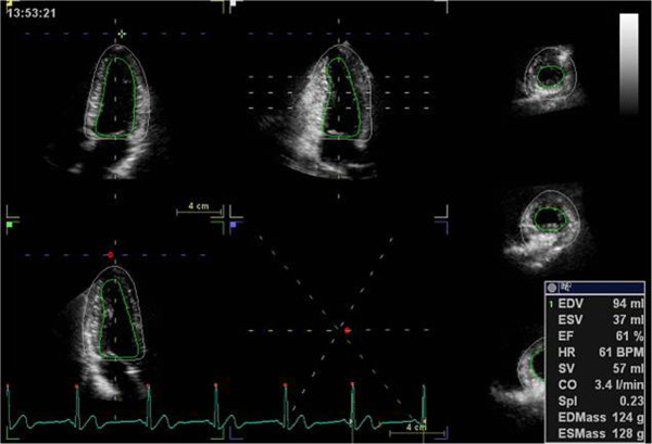
Background: Pregnancy represents a physiological adaptation to the transient load changes of maternal heart. This study aimed to investigate maternal left ventricle (LV) performance during normal pregnancy by three-dimensional speckle-tracking echocardiography (3D STE) parameters considering LV loading and shape.

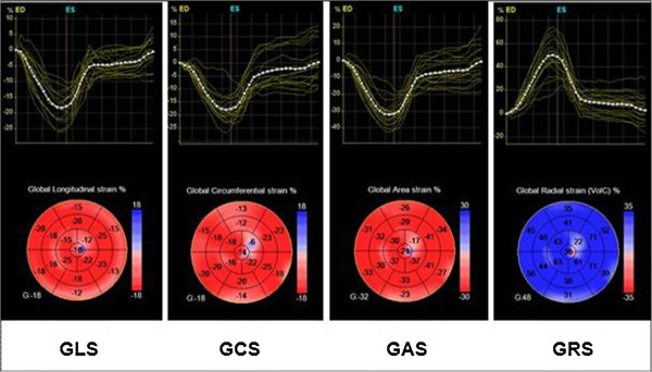
Methods: Sequential two-dimensional echocardiography (2DE) and 3D STE were performed on 68 women during each pregnancy trimester and 6 to 9 weeks after delivery, while thirty age-matched, healthy, nonpregnant women served as controls. Global longitudinal strain (GLS), global circumferential strain (GCS), global area strain (GAS) and global radial strain (GRS) were measured.

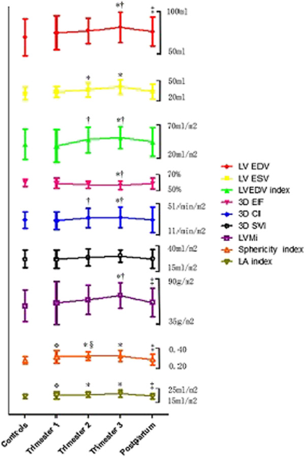
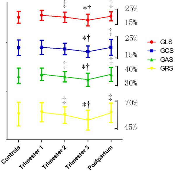
Results: Increased cardiac index and progressive eccentric hypertrophy was detected, which subsequently recovered postpartum. In late pregnancy, GLS, GCS, GAS and GRS significantly decreased (P < 0.05) accompanied by a slight reduction of LV ejection fraction (EF) (P < 0.05), and these values returned postpartum to baseline level. All 3D strain indices correlated well with gestation age (P < 0.01), while compared to other components, GAS exhibited the strongest association with 3D EF (r = 0.549) and sphericity index (r = 0.328), and was the only parameter that correlated well with LV mass index (r = 0.22).

Conclusions: This study gives normal ranges of 3D STE indices in pregnancy. 3D STE demonstrated modified myocardial deformation and changes in maternal LV structure and function during the gestation period.


Figures

Figure 1
Figure 1
Automated detection of the endocardium and epicardial border throughout the whole cardiac cycle. Consequently end-diastolic volume (EDV), end-systolic volume (ESV), ejection fraction (EF), stroke volume (SV), cardiac output (CO), sphericity index(Spl) and LV mass either at end-diastole (EDMass) or at end-systole (ESMass) were automatically calculated.
Figure 2
Figure 2
Both strain curves and a color-coded 17-segment bull’s eye plot were presented. Color lines indicate regional strain; white dotted line means global (average) strain. Values of longitudinal strain, circumferential strain, and area strain are negative (sign -), whereas values of radial strain are positive (sign +). GAS indicates global area strain; GCS, global circumferential strain; GLS, global longitudinal strain; GRS, global radial strain.
Figure 3
Figure 3
Evolution of maternal LV morphology and function during pregnancy and postpartum by 3D STE. Data are presented as mean ± SD. LVEDV indicates left ventricular end-diastolic volume; LVESV, left ventricular end-systolic volume; LVEDVi, left ventricular end-diastolic volume index; EF, ejection fraction; CI, cardiac index; SVI, stroke volume index; LVMi, left ventricular mass index; LA index, left atrial volume index; *P < 0.05 vs. Controls; †P < 0.05 vs. Trimester 1; ‡P < 0.05 vs. Trimester 3; §P < 0.05 vs. Postpartum.
Figure 4
Figure 4
Assessment of myocardial deformation during pregnancy and postpartum by 3D STE. Data are presented as mean ± SD. Note that GLS, GCS, GRS and GAS decreased in Trimester 3, while returned in postpartum. GLS indicates global longitudinal strain; GCS, global circumferential strain; GAS, global area strain; GRS, global radial strain. *P < 0.05 vs. Controls; †P < 0.05 vs. Trimester 1; ‡P < 0.05 vs. Trimester 3; §P < 0.05 vs. Postpartum.

References

    1. Presbitero P, Boccuzzi GG, Groot CJM, Roos-Hesselink JW. ESC textbook of cardiovascular medicine. Oxford: Oxford University Press; 2009.
    1. Siu SC, Sermer M, Colman JM, Alvarez AN, Mercier LA, Morton BC, et al. Prospective multicenter study of pregnancy outcomes in women with heart disease. Circulation. 2001;104:515–21. doi: 10.1161/hc3001.093437. - DOI - PubMed
    1. Desai DK, Moodley J, Naidoo DP. Echocardiographic assessment of cardiovascular hemodynamics in normal pregnancy. Obstet Gynecol. 2004;104:20–9. doi: 10.1097/01.AOG.0000128170.15161.1d. - DOI - PubMed
    1. Simmons LA, Gillin AG, Jeremy RW. Structural and functional changes in left ventricle during normotensive and preeclamptic pregnancy. Am J Physiol Heart Circ Physiol. 2002;283:H1627–33. doi: 10.1152/ajpheart.00966.2001. - DOI - PubMed
    1. Bamfo JE, Kametas NA, Nicolaides KH, Chambers JB. Reference ranges for tissue Doppler measures of maternal systolic and diastolic left ventricular function. Ultrasound Obstet Gynecol. 2007;29:414–20. doi: 10.1002/uog.3966. - DOI - PubMed

Publication types