. 2015 Jun;96(Pt 6):1411-1422.
doi: 10.1099/vir.0.000071. Epub 2015 Jan 27.

Discovery of a polyomavirus in European badgers (Meles meles) and the evolution of host range in the family Polyomaviridae

Affiliations

Discovery of a polyomavirus in European badgers (Meles meles) and the evolution of host range in the family Polyomaviridae

Sarah C Hill et al. J Gen Virol. 2015 Jun.

Abstract

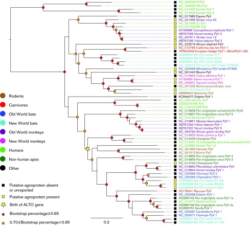
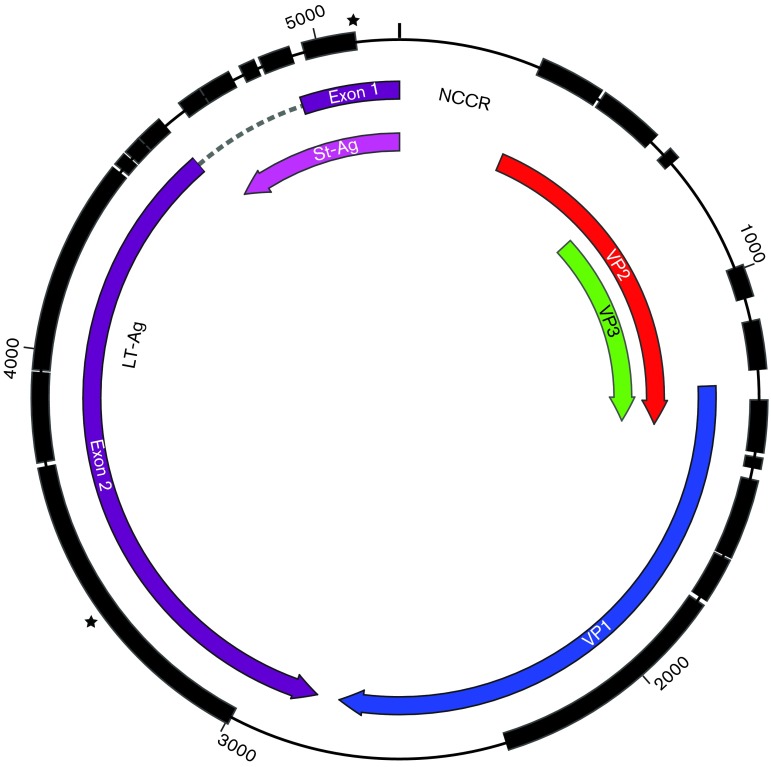
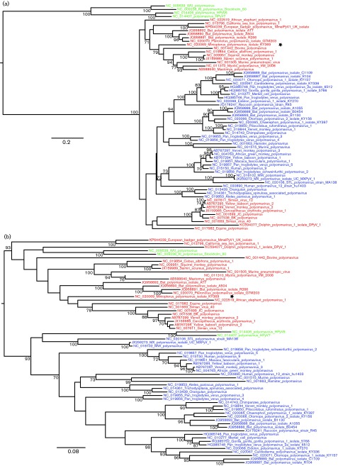
Polyomaviruses infect a diverse range of mammalian and avian hosts, and are associated with a variety of symptoms. However, it is unknown whether the viruses are found in all mammalian families and the evolutionary history of the polyomaviruses is still unclear. Here, we report the discovery of a novel polyomavirus in the European badger (Meles meles), which to our knowledge represents the first polyomavirus to be characterized in the family Mustelidae, and within a European carnivoran. Although the virus was discovered serendipitously in the supernatant of a cell culture inoculated with badger material, we subsequently confirmed its presence in wild badgers. The European badger polyomavirus was tentatively named Meles meles polyomavirus 1 (MmelPyV1). The genome is 5187 bp long and encodes proteins typical of polyomaviruses. Phylogenetic analyses including all known polyomavirus genomes consistently group MmelPyV1 with California sea lion polyomavirus 1 across all regions of the genome. Further evolutionary analyses revealed phylogenetic discordance amongst polyomavirus genome regions, possibly arising from evolutionary rate heterogeneity, and a complex association between polyomavirus phylogeny and host taxonomic groups.


Figures

Fig. 1.
Fig. 1.. Maximum-likelihood phylogeny of all known polyomavirus (PyV) species or putative species, including MmelPyV1, estimated from the ‘genome-wide’ alignment. Viral taxa are coloured according to host species and bootstrap support scores are indicated using coloured circles (as indicated in the key). Scale bar represents expected number of substitutions per site (first and second codon positions only).
Fig. 2.
Fig. 2.. Genome map of MmelPyV1. Thick black blocks (outer circle) represent genomic regions that could be aligned across all mammalian polyomaviruses and were therefore included in the ‘genome-wide’ alignment. Coloured arrows represent ORFs. Early proteins are in purple; late proteins are in blue, red and green. Breakpoints used in recombination analysis are marked with two stars.
Fig. 3.
Fig. 3.. Gel electrophoresis of mink cell line NBL-7. Lanes: I, infected with UK isolate cell line; M, mock infected. Numbers below the lanes represent the primers used. Numbers on the left represent the ladder fragment size (bp). Controls not shown.
Fig. 4.
Fig. 4.. Conserved ORFs of the two MmelPyV1 isolates and their nearest relative (CSLPyV1). All six translation frames are shown; the three forward (For) and reverse (Rev) reading frames are shown in separate boxes. All ORFs >100 aa are shown as boxes, with the first methionine marked in red. Solid vertical lines indicate stop codons and tick marks indicate 1000 bp markers. Blue coloured ORFs represent probable protein-coding ORFs identifiable by numbering: 1, VP2 and VP3; 2, VP1; 3, St-Ag and exon 1 of LT-Ag; 4, exon 2 of LT-Ag. Details of GenBank accession numbers and genome length are also included.
Fig. 5.
Fig. 5.. Maximum-likelihood phylogenies produced from different regions of the genome. (a) Partition A, comprising the late region plus a short region of LT-Ag exon 2. (b) Partition B, comprising the majority of the early region. Red and blue coloured branches indicate the major two clades as identified in the genome-wide alignment (Fig. 1). Green branches indicate human polyomaviruses within inconsistent phylogenetic locations. The Miniopterus polyomavirus is marked with a star to indicate identification in Recco as a further possible recombinant. Scale bar represents expected number of substitutions per site (first and second codon positions only).

References

    1. An P., Sáenz Robles M. T., Pipas J. M. ( 2012. ). Large T antigens of polyomaviruses: amazing molecular machines. Annu Rev Microbiol 66, 213–236. 10.1146/annurev-micro-092611-150154 - DOI - PubMed
    1. Baker T. S., Drak J., Bina M. ( 1989. ). The capsid of small papova viruses contains 72 pentameric capsomeres: direct evidence from cryo-electron-microscopy of simian virus 40. Biophys J 55, 243–253. 10.1016/S0006-3495(89)82799-7 - DOI - PMC - PubMed
    1. Banks M., King D. P., Daniells C., Stagg D. A., Gavier-Widen D. ( 2002. ). Partial characterization of a novel gammaherpesvirus isolated from a European badger (Meles meles). J Gen Virol 83, 1325–1330. - PubMed
    1. Carter J. J., Daugherty M. D., Qi X., Bheda-Malge A., Wipf G. C., Robinson K., Roman A., Malik H. S., Galloway D. A. ( 2013. ). Identification of an overprinting gene in Merkel cell polyomavirus provides evolutionary insight into the birth of viral genes. Proc Natl Acad Sci U S A 110, 12744–12749. 10.1073/pnas.1303526110 - DOI - PMC - PubMed
    1. Charleston M. A., Robertson D. L. ( 2002. ). Preferential host switching by primate lentiviruses can account for phylogenetic similarity with the primate phylogeny. Syst Biol 51, 528–535. 10.1080/10635150290069940 - DOI - PubMed

Publication types

MeSH terms

Associated data

LinkOut - more resources