Review
. 2015 Apr 22;4(6):811-30.
doi: 10.1002/adhm.201400715. Epub 2015 Jan 27.

Spatio-Temporal Control of LbL Films for Biomedical Applications: From 2D to 3D

Affiliations
Review

Spatio-Temporal Control of LbL Films for Biomedical Applications: From 2D to 3D

Claire Monge et al. Adv Healthc Mater. .

Abstract

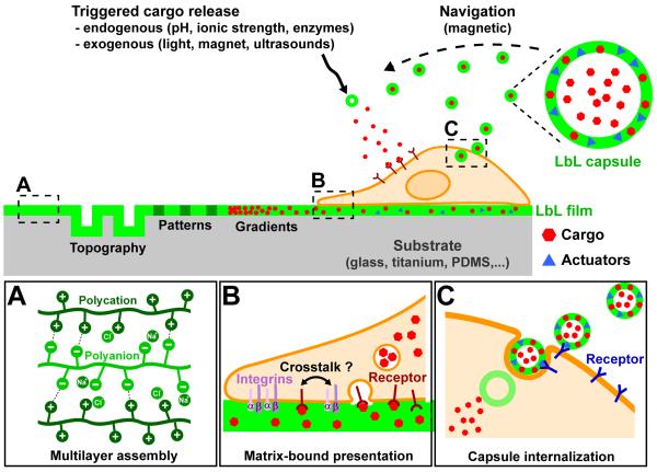
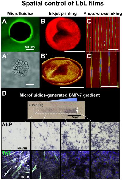
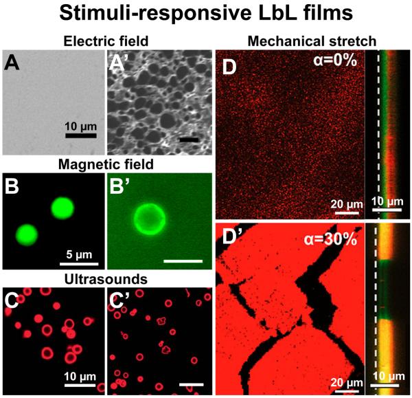
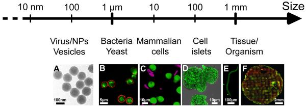
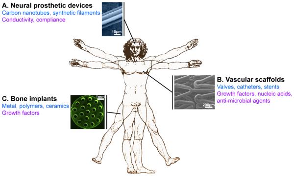
Introduced in the '90s by Prof. Moehwald, Lvov, and Decher, the layer-by-layer (LbL) assembly of polyelectrolytes has become a popular technique to engineer various types of objects such as films, capsules and free standing membranes, with an unprecedented control at the nanometer and micrometer scales. The LbL technique allows to engineer biofunctional surface coatings, which may be dedicated to biomedical applications in vivo but also to fundamental studies and diagnosis in vitro. Initially mostly developed as 2D coatings and hollow capsules, the range of complex objects created by the LbL technique has greatly expanded in the past 10 years. In this Review, the aim is to highlight the recent progress in the field of LbL films for biomedical applications and to discuss the various ways to spatially and temporally control the biochemical and mechanical properties of multilayers. In particular, three major developments of LbL films are discussed: 1) the new methods and templates to engineer LbL films and control cellular processes from adhesion to differentiation, 2) the major ways to achieve temporal control by chemical, biological and physical triggers and, 3) the combinations of LbL technique, cells and scaffolds for repairing 3D tissues, including cardio-vascular devices, bone implants and neuro-prosthetic devices.

Keywords: drug delivery; layer-by-layer; scaffolds; spatial; temporal; tissue engineering.


Figures

FIGURE 1
FIGURE 1. A schematic overview of the use of LbL films and microcapsules to influence cell behavior
The left part illustrates the possibilities of spatially controlling LbL films to present a structured topography, patterns and gradients of rigidity or bioactivity. The right part illustrates the possibilities of loading in LbL films or capsules a cargo of interest (e.g. proteins, drugs, siRNA,…) to be delivered to the cell, as well as actuators (e.g. antibodies, gold NPs, magnetic NPs,…). These actuators can be used for targeting specific cell types or triggering the cargo release. The boxes (A - C) zoom in on areas of interest. (A) Electrostatic interactions in LbL films. Representation of the electrostatic interactions between polycations and polyanions and the types of charge compensation in a LbL assembly. (B) Presentation of a cargo by LbL films. This matrix-bound presentation allows spatial confinement of adhesion receptors (e.g. integrins, represented here by their α and β sub-units) and cargo receptors at the ventral side of the cell. Due to the close proximity of cargo receptors and adhesion receptors, a crosstalk between these two types of receptor is possible. The internalization of the cargo by endocytosis is also illustrated. (C) Presentation of a cargo by LbL capsules. The use of specific actuators (antibodies, glycans, folates, hyaluronic acid) allows the delivery of a loaded cargo to a designated cell type (e.g. tumor cells). The LbL capsules can be internalized by endocytosis in the cell.
FIGURE 2
FIGURE 2. Illustrations of recent developments in spatially controlled LbL films
(A) Fluorescence image and (A’) phase-contrast of the selective deposition by microfluidics of a poly-L-lysine/hyaluronic acid (PLL/HA) film inducing selective growth of L929 cells in the uncoated spherical areas. (B) Fluorescence image and (B’) 3D AFM image of a rhodamine-loaded multilayer disc assembled by inkjet printing. (C) Overview and (C’) magnification of the orientation of C2C12 myoblasts, stained for vinculin (green), actin (red) and nucleus (blue), on linear micropatterns of rigidity engineered by photo-crosslinking of a PLL/HA film. Red background corresponds to the autofluorescence of the chromium mask. (D) Differentiation of C2C12 myoblasts on a PLL/HA film presenting a matrix-bound bone morphogenetic protein-7 (BMP-7) gradient generated by microfluidics. Overview image and representative images of alizarin red (ALP) staining (dark purple) confirms osteogenic differentiation, while immunofluorescent imaging reveals a decrease of troponin T (TnT, green) positive cells with increasing BMP-7 concentration. Actin is stained in gray and nuclei in blue. A and A’ adapted from [20] with permission of The Royal Society of Chemistry, B and B’ from [33] with permission of the American Chemical Society, C, C’ and D from [60] and [66] with permission of Elsevier.
FIGURE 3
FIGURE 3. Illustrations of recent developments in stimuli-responsive LbL films
(A) SEM images (top-view) of LPEI/PAA LbL films before and (A’) after 1h exposure to an electric field, inducing a strong change of the film permeability. (B) Fluorescence images of hybrid capsules formed with PSS/PAH, Fe3O4 nanoparticles and a DDAC bilayer membrane loaded with calcein before and (B’) after alternating magnetic field irradiation at 360 kHz and 234 Oe for 30 min, inducing the immediate release of the calcein. (C) Confocal images of BSARhodamine-loaded microcapsules of PSS/PAH before and (C’) after ultrasonic irradiation at 3.19 W for 5 min, inducing some partial and full ruptures of the microcapsules. (D) Confocal images of a (PLL/HA)/(PAH/PSS) incubated with trypsinRhodamine solution in a non-stretched and (D’) a 30% stretch state, inducing trypsinRhodamine diffusion across the PAH/PSS barrier and within the PLL/HA reservoir. Left images were acquired in (x,y) plane whereas right images were acquired in (x,z) plane at the reservoir–barrier interface. The white dashed line indicates the film-silicone substrate interface. A, A’, B, B’, D and D’ adapted from [133], [142] and [155] with permission of the American Chemical Society. C and C’ adapted from [149] with permission of The Royal Society of Chemistry.
FIGURE 4
FIGURE 4. LbL coatings of 3D objects over a wide range of sizes
Recent uses of the LbL technique to coat 3D objects from nanometric objects to millimer size tissues or organisms: A). polystyrene latex NPs, B) yeast, e.g. yEGFP-expressing S. cerevisiae, C) mammalian cells e.g. monocytes, D) cell islets e.g. pancreatic islets, E) organisms, e.g. nematodes C. elegans and F) millimetric engineered tissues, e.g. 3D tissue made of human dermal fibroblasts and HUVECs. A, C, D and E adapted from [106], [30], [178] and [245] with permission of the American Chemical Society. B adapted from [246] with permission of The Royal Society of Chemistry. F adapted from [247] with permission of Elsevier.
FIGURE 5
FIGURE 5. Methods for the production of LbL freestanding membranes made of polyelectrolytes or living cells
(A) The LbL films can be released from the substrate by dissolution of a sacrificial layer, (B) by peeling off the substrate using a dissolvable supporting layer or (C) by direct in case of low interactions between the LbL film and the underlying substrate. (D) A freestanding cell sheet can be produced by dissolution of a sacrificial LbL film, on top of which cells are forming a sheet. These freestanding membranes can be used for tissue engineering and repair. A, B and C adapted from [191], [194] and [197] with permission of the American Chemical Society. D adapted from [200] with permission of Elsevier.
FIGURE 6
FIGURE 6. LbL coating for 3D scaffolds in vivo
Three main medical applications are concerned by LbL functional coatings. (A) On neural prosthetic devices, the LbL coating provides conductivity and compliance to the carbon nanotubes or synthetic filaments (illustration of a neural electrode). (B) Vascular replacement or repair is approached by coating decellularized matrices, catheters or stents that deliver growth factors (VEGF), nucleic acids (SiRNA or plasmid DNA) or antimicrobial agents (β-peptide, Nitric oxide, cateslytin…) (illustration of a LbL-coated stent). (C) Bone regeneration is addressed by the delivery of BMP-2 from LbL-coated metal, polymer or ceramic implants (illustration of a titanium bone implant). In black: medical field; in blue: scaffolds used; in purple: biochemical, physical or mechanical signals provided by the LbL coating. A and C adapted from [207] and [242] with permission of Elsevier. B adapted from [216] with the permission of the American Chemical Society.

References

    1. Takahashi H, Letourneur D, Grainger DW. Biomacromolecules. 2007;8:3281. - PMC - PubMed
    1. Farhatnia Y, Tan A, Motiwala A, Cousins BG, Seifalian AM. Biotechnol. Adv. 2013;31:524. - PubMed
    1. Decher G. Science. 1997;277:1232. (80-. )
    1. Moya S, Sukhorukov G, Auch M, Donath E, Möhwald H. J. Colloid Interface Sci. 1999;216:297. - PubMed
    1. Lvov Y, Onda M, Ariga K, Kunitake T. J. Biomater. Sci. Polym. Ed. 1998;9:345. - PubMed

Publication types

LinkOut - more resources