. 2015 Mar 13;290(11):6951-61.
doi: 10.1074/jbc.M114.606186. Epub 2015 Jan 27.

Myofibroblasts exhibit enhanced fibronectin assembly that is intrinsic to their contractile phenotype

Affiliations

Myofibroblasts exhibit enhanced fibronectin assembly that is intrinsic to their contractile phenotype

Elizabeth E Torr et al. J Biol Chem. .

Abstract

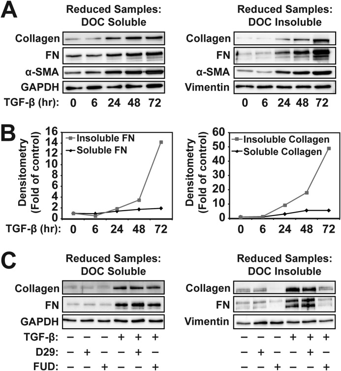
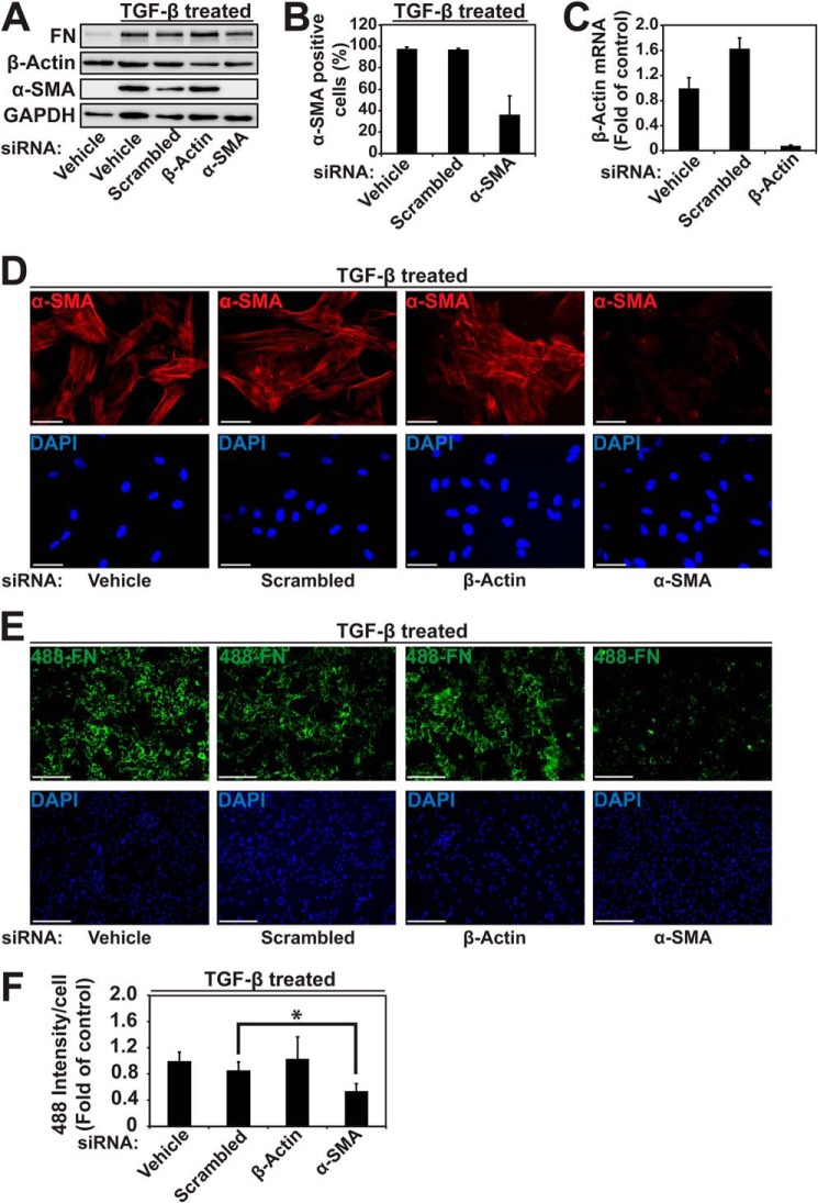
Myofibroblasts have increased expression of contractile proteins and display augmented contractility. It is not known if the augmented contractile gene expression characterizing the myofibroblast phenotype impacts its intrinsic ability to assemble fibronectin (FN) and extracellular matrix. In this study we investigated whether myofibroblasts displayed increased rates of FN fibril assembly when compared with their undifferentiated counterparts. Freshly plated myofibroblasts assemble exogenous FN (488-FN) into a fibrillar matrix more rapidly than fibroblasts that have not undergone myofibroblast differentiation. The augmented rate of FN matrix formation by myofibroblasts was dependent on intact Rho/Rho kinase (ROCK) and myosin signals inasmuch as treatment with Y27632 or blebbistatin attenuated 488-FN assembly. Inhibiting contractile gene expression by pharmacologic disruption of the transcription factors megakaryoblastic leukemia-1 (MKL1)/serum response factor (SRF) during myofibroblast differentiation resulted in decreased contractile force generation and attenuated 488-FN incorporation although not FN expression. Furthermore, disruption of the MKL1/SRF target gene, smooth muscle α-actin (α-SMA) via siRNA knockdown resulted in attenuation of 488-FN assembly. In conclusion, this study demonstrates a linkage between increased contractile gene expression, most importantly α-SMA, and the intrinsic capacity of myofibroblasts to assemble exogenous FN into fibrillar extracellular matrix.

Keywords: Extracellular Matrix; Fibroblast; Fibronectin; Megakaryoblastic Leukemia-1; Myofibroblast; Serum Response Factor; Smooth Muscle α-Actin; Transforming Growth Factor β (TGF-B).


Figures

FIGURE 1.
FIGURE 1.
Collagen deposition into the extracellular matrix accelerates during myofibroblast differentiation and requires fibrillar fibronectin assembly. HLF were treated with 1 ng/ml TGF-β for the times indicated. A, cells were extracted with a 2% DOC-containing buffer and reduced DOC-soluble and -insoluble lysates subjected to PAGE and immunoblotted with the indicated antibodies. B, densitometry of Western blots of the indicated lysates and proteins. Data are normalized to the control condition for each protein to compare kinetics. C, HLF were treated with 1 ng/ml TGF-β for 24 h in the presence of the 56-residue FUD of the F1 adhesin protein of S. pyogenes (500 nm), D29 peptide control (500 nm), or vehicle alone. Cells were subjected to DOC extraction, with reduced samples subjected to PAGE and Western blotting with the indicated antibodies.
FIGURE 2.
FIGURE 2.
Differentiated myofibroblasts retain their phenotype but lose endogenous FN expression upon re-plating in the presence of cycloheximide. HLF were treated with or without 1 ng/ml TGF-β for 72 h to induce a myofibroblast phenotype. After differentiation, cells were then trypsinized and re-plated in the presence or absence of 10 μm cycloheximide. A, after 6 h of attachment, cells were lysed with radioimmune precipitation lysate and subjected to PAGE and immunoblotting with the indicated antibodies. B and C, fibroblasts (untreated) or myofibroblasts (treated with TGF-β for 72 h) were trypsinized and re-plated in the presence of 10 μm cycloheximide and allowed to attach and spread for 6 h. Cells were then fixed, and immunofluorescent staining was performed against α-SMA (B) or endogenous FN (C). Cells were counterstained with DAPI (scale bar = 250 μm).
FIGURE 3.
FIGURE 3.
Myofibroblasts assemble exogenous fibronectin more rapidly than undifferentiated fibroblasts. Fibroblasts and myofibroblasts (treated with 1 ng/ml TGF-β for 72 h) were trypsinized and re-plated in the presence of 10 μm cycloheximide as in Fig. 2. After 6 h of attachment, they were then incubated with 4 μg/ml 488-FN for 0.5, 1.5, 6, or 24 h followed by fixation and counterstained with DAPI. A, integrated intensity of the 488 signal was quantified using ImageJ. Student's t test with Bonferroni correction for multiple tests (total allowable false discovery rate = 0.05, thus *, p ≤ 0.05/4 = 0.0125) was used for statistical analysis. NS, not significant. B, representative images from a 1.5-h incubation with 488-FN, with high power images showing the fibrillar matrix (scale bar = 250 μm for the top four panels and 50 μm for the bottom two panels). C, representative images from 24 h of incubation with 488-FN (scale bar = 250 μm). D, incubation with 4 μg/ml 488-FN for 6 h followed by DOC extraction and fixation showing residual DOC-insoluble matrix. Cells are absent due to DOC extraction (scale bar = 250 μm).
FIGURE 4.
FIGURE 4.
Colchicine-induced myofibroblasts assemble exogenous fibronectin more rapidly than undifferentiated fibroblasts. A, treatment of HLF with 1 μm colchicine (colch) for 24 h results in induction of α-SMA. B, fibroblasts and myofibroblasts (treated with 1 μm colchicine for 24 h) were trypsinized and re-plated in the presence of 10 μm cycloheximide, as in Fig. 2. After 6 h of attachment, they were then incubated with 4 μg/ml 488-FN for 1.5 h followed by fixation and counterstained with DAPI. Quantification of 488-FN incorporation using ImageJ was as in Fig. 3A. Student's t test (*, p < 0.05) was used for statistical analysis. C, representative images after 1.5 h of incubation with 488-FN after fixation and DAPI co-stain (scale bar = 250 μm).
FIGURE 5.
FIGURE 5.
Augmented fibronectin assembly by myofibroblasts requires ROCK and myosin. A, after induction of the myofibroblast phenotype by 72 h of treatment with TGF-B, cells were then treated with 10 μm Y27632 (0.5 or 2 h), 50 μm blebbistatin (0.5 h), or vehicle control. In one condition, 10 μm Y27632 was added during the differentiation phase (0.5 h before treatment with TGF-β, 72.5 h total). Whole cell lysates were then subjected to PAGE followed by immunoblotting with the indicated antibodies. B, phospho-cofilin/GAPDH and phospho-MLC/GAPDH signals were quantified using ImageQuant software and normalized to TGF-β-stimulated controls. Student's t test with Bonferroni correction (n = 2, thus *, p < 0.025) was used for statistical analysis. C, cells were differentiated in the presence or absence of 1 ng/ml TGF-β for 72 h followed by re-plating as in Fig. 3. After 6 h of attachment, inhibitors were added for 0.5 h, and then cells were incubated with 4 μg/ml 488-FN for 1.5 h, fixed, and counterstained with DAPI. Quantification of the 488-FN incorporation was then performed using ImageJ. Student's t test with Bonferroni correction (n = 2, thus *, p < 0.025) was used for statistical analysis. D, representative images from the 1.5-h incubation with 488-FN (scale bar = 250 μm).
FIGURE 6.
FIGURE 6.
MKL1/SRF is required for maintenance of the contractile phenotype. A, HLF were co-transfected with cDNAs for the SRF firefly luciferase reporter, SRE.L, Renilla luciferase driven by thymidine kinase promoter, together with either empty vector (pcDNA3.1) or cDNA for full-length MKL1. After transfection, cells were incubated overnight in serum-free media in the presence or absence of 10 μm CCG-1423. Luciferase activity was measured as described under “Experimental Procedures.” Student's t test (*, p < 0.05) was used for statistical comparison of vehicle and CCG-treated conditions. B, HLF were co-transfected with cDNAs for Smad-binding element (SBE)-driven firefly luciferase and Renilla luciferase driven by thymidine kinase promoter. After serum starvation, cells were incubated with 10 μm CCG-1423 (or vehicle) for 0.5 h followed by stimulation with 1 ng/ml TGF-β for 6 h. Cell lysates were run for luciferase activity. Student's t test (p <0.05) was used for statistical comparison of vehicle and CCG-treated conditions. NS, not significant. C, HLF were treated with 1 ng/ml TGF-β for 72 h in the presence of 10 μm CCG-1423 or vehicle control. Cells were seeded into collagen gels, released, and observed for contraction. Digital images were taken 48 h after gel release. D, gel areas were quantified from the digital images at each time point using ImageJ. Student's t test (*, p < 0.05) was used for statistical comparison of TGF-β and TGF-β/CCG groups 48 h post-release.
FIGURE 7.
FIGURE 7.
Fibronectin incorporation in myofibroblasts requires intact MKL1 gene expression. A, HLF were stimulated with 1 ng/ml TGF-β1 in the presence of 10 μm CCG-1423 or vehicle control (added 30 min before stimulation) for 24 h. Cell lysates were separated into DOC-soluble and -insoluble fractions that were then treated with reducing sample buffers. Lysates were then subjected to PAGE on an 8% gel followed by immunoblotting with antibodies against total fibronectin, GAPDH, and vimentin. Fibronectin antibodies detect two predominant isoforms of fibronectin that are resolvable on 8% gels. B, cells were stimulated with 1 ng/ml TGF-β in the presence or absence of 10 μm CCG-1423 (added 30 min before stimulation) for 24 h. Whole cell lysates (in radioimmune precipitation assay buffer) were analyzed by PAGE on a 10% gel followed by immunoblotting with the indicated. C, HLF were stimulated with 1 ng/ml TGF-β for 72 h in the presence of 10 μm CCG-1423 or vehicle control (added 30 min before stimulation). After differentiation, cells were re-plated in the presence of cycloheximide and allowed to adhere for 6 h (as in Figs. 2 and 3). 488-FN at 4 μg/ml was then added for 1.5 h followed by fixation and co-staining with DAPI (scale bar = 125 μm). D, quantitation of 488-FN incorporation using ImageJ. Student's t test (*, p < 0.05) was used for statistical analysis. E, HLF were differentiated with 1 ng/ml TGF-β for 72 h followed by re-plating and a 6-h attachment as above. After attachment, cells were then incubated with 10 μm CCG-1423 or vehicle control 30 min before the addition of 488-FN. Incubation with 4 μg/ml 488-FN for 1.5 h was followed by fixation and counterstained with DAPI (scale bar = 60 μm). F, quantification of 488-FN incorporation was done using ImageJ. Student's t test (p < 0.05) was used for statistical analysis. NS, not significant.
FIGURE 8.
FIGURE 8.
Smooth muscle α-actin expression is required for fibronectin assembly by myofibroblasts. HLF were transfected with scrambled siRNA, siRNA against β-actin, or siRNA against α-SMA and then treated with 1 ng/ml TGF-β for 72 h. A, cell lysates were subjected to PAGE and immunoblotting for the indicated antibodies. B, HLF treated as above were also fixed and stained for α-SMA and DAPI. α-SMA-positive cells were manually counted for 10 high power fields for the indicated conditions. C, HLF were transfected with siRNA against β-actin or controls as indicated. Lysates were analyzed for β-actin mRNA expression using quantitative real time PCR. D and E, HLF transfected with scrambled siRNA, siRNA against β-actin, or siRNA against α-SMA and then treated with 1 ng/ml TGF-β for 72 h were re-plated and allowed to attach for 6 h in the presence of 10 μm cycloheximide as in Figs. 2 and 3. Cells were then either fixed and co-stained for α-SMA (scale bar = 250 μm) (D) or incubated with 4 μg/ml 488-FN for 1.5 h (E) followed by fixation and imaging. F, quantification of 488-FN assembly using ImageJ. Student's t test (*, p < 0.05) was used for statistical analysis.

Similar articles

Cited by

References

    1. Phan S. H. (1996) Role of the myofibroblast in pulmonary fibrosis. Kidney Int. Suppl. 54, S46–S48 - PubMed
    1. Gabbiani G., Ryan G. B., Majne G. (1971) Presence of modified fibroblasts in granulation tissue and their possible role in wound contraction. Experientia 27, 549–550 - PubMed
    1. Malmström J., Lindberg H., Lindberg C., Bratt C., Wieslander E., Delander E. L., Särnstrand B., Burns J. S., Mose-Larsen P., Fey S., Marko-Varga G. (2004) Transforming growth factor-β1 specifically induce proteins involved in the myofibroblast contractile apparatus. Mol. Cell. Proteomics 3, 466–477 - PubMed
    1. Sottile J., Hocking D. C. (2002) Fibronectin polymerization regulates the composition and stability of extracellular matrix fibrils and cell-matrix adhesions. Mol. Biol. Cell 13, 3546–3559 - PMC - PubMed
    1. Sabatier L., Chen D., Fagotto-Kaufmann C., Hubmacher D., McKee M. D., Annis D. S., Mosher D. F., Reinhardt D. P. (2009) Fibrillin assembly requires fibronectin. Mol. Biol. Cell 20, 846–858 - PMC - PubMed

Publication types